Institute of Cellular Neurosciences, University of Bonn, Sigmund Freud Strasse 25, Bonn, Germany.
Epilepsia. 2012 Nov;53(11):1898-906. doi: 10.1111/j.1528-1167.2012.03665.x. Epub 2012 Sep 11.
Dysfunction of the blood-brain barrier (BBB) and albumin extravasation have been suggested to play a role in the etiology of human epilepsy. In this context, dysfunction of glial cells attracts increasing attention. Our study was aimed to analyze in the hippocampus (1) which cell types internalize albumin injected into the lateral ventricle in vivo, (2) whether internalization into astrocytes impacts their coupling and expression of connexin 43 (Cx43), and (3) whether expression of Kir4.1, the predominating astrocytic K(+) channel subunit, is altered by albumin.
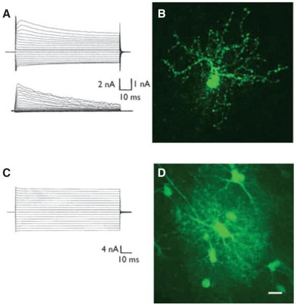
The patch-clamp method was combined with single cell tracer filling to investigate electrophysiologic properties and gap junction coupling (GJC). For cell identification, mice with cell type-specific expression of a fluorescent protein (NG2kiEYFP mice) and immunohistochemistry were employed. Semiquantitative real time polymerase chain reaction (RT-PCR) allowed analysis of Kir4.1 and Cx43 transcript levels.
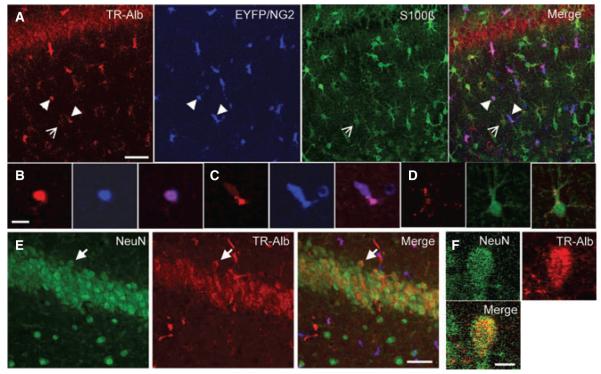
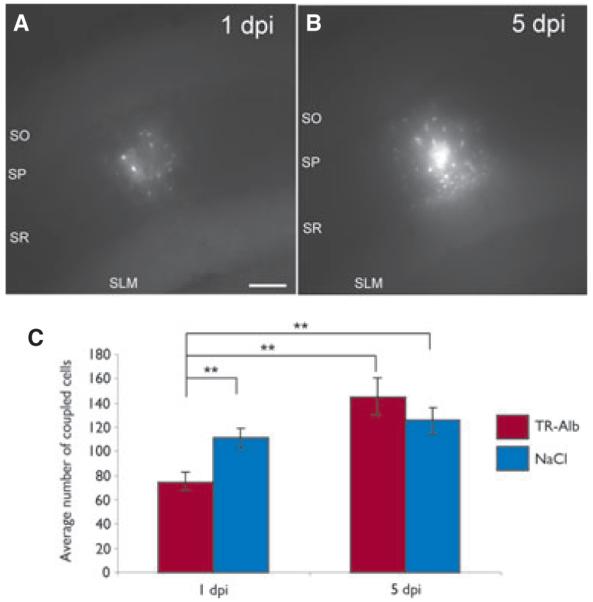
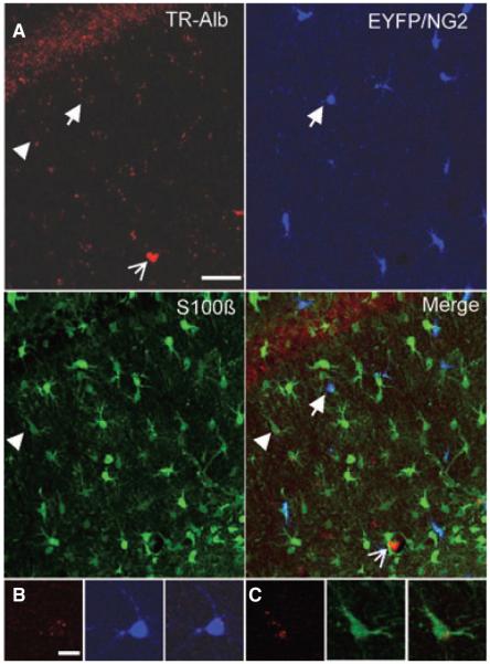
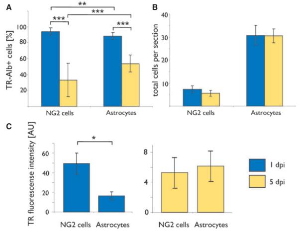
We show that fluorescently labeled albumin is taken up by astrocytes, NG2 cells, and neurons, with NG2 cells standing out in terms of the quantity of uptake. Within 5 days postinjection (dpi), intracellular albumin accumulation was largely reduced suggesting rapid degradation. Electrophysiologic analysis of astrocytes and NG2 cells revealed no changes in their membrane properties at either time point. However, astrocytic GJC was significantly decreased at 1 dpi but returned to control levels within 5 dpi. We found no changes in hippocampal Cx43 transcript expression, suggesting that other mechanisms account for the observed changes in coupling. Kir4.1 mRNA was regulated oppositely in the CA1 stratum radiatum, with a strong increase at 1 dpi followed by a decrease at 5 dpi.
The present study demonstrates that extravasal albumin in the hippocampus induces rapid changes of astrocyte function, which can be expected to impair ion and transmitter homeostasis and contribute to hyperactivity and epileptogenesis. Therefore, astrocytes may represent alternative targets for antiepileptic therapeutic approaches.
血脑屏障(BBB)功能障碍和白蛋白渗出被认为在人类癫痫的发病机制中起作用。在这种情况下,神经胶质细胞的功能障碍引起了越来越多的关注。我们的研究旨在分析海马体中:(1)哪种细胞类型会将注入侧脑室的白蛋白内化;(2)白蛋白内化是否会影响星形胶质细胞的偶联和连接蛋白 43(Cx43)的表达;(3)Kir4.1(主要的星形胶质细胞 K+通道亚基)的表达是否会因白蛋白而改变。
我们将膜片钳技术与单细胞示踪剂填充相结合,以研究电生理特性和缝隙连接偶联(GJC)。为了细胞鉴定,我们使用了具有荧光蛋白(NG2kiEYFP 小鼠)细胞类型特异性表达的小鼠和免疫组织化学方法。半定量实时聚合酶链反应(RT-PCR)允许分析 Kir4.1 和 Cx43 的转录水平。
我们表明,荧光标记的白蛋白被星形胶质细胞、NG2 细胞和神经元摄取,其中 NG2 细胞在摄取量方面表现突出。在注射后 5 天(dpi)内,细胞内白蛋白的积累量大大减少,表明其快速降解。在两个时间点,对星形胶质细胞和 NG2 细胞的电生理分析都没有发现其膜特性的变化。然而,星形胶质细胞的 GJC 在 1dpi 时显著降低,但在 5dpi 时恢复到对照水平。我们没有发现海马 Cx43 转录表达的变化,这表明其他机制解释了偶联的变化。Kir4.1mRNA 的调节在 CA1 放射状层中相反,在 1dpi 时强烈增加,然后在 5dpi 时减少。
本研究表明,海马体中的外渗白蛋白会引起星形胶质细胞功能的快速变化,这可能会损害离子和递质的稳态,并导致过度兴奋和癫痫发生。因此,星形胶质细胞可能是抗癫痫治疗方法的替代靶点。