Department of Surgery, Eulji Medical College Hospital, Seoul, Korea.
J Korean Med Sci. 2012 Sep;27(9):1019-26. doi: 10.3346/jkms.2012.27.9.1019. Epub 2012 Aug 22.
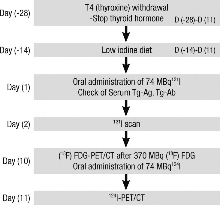
Although the prognosis of patients with differentiated thyroid carcinoma (DTC) is generally encouraging, a diagnostic dilemma is posed when an increasing level of serum thyroglobulin (Tg) is noted, without detection of a recurrent tumor using conventional imaging tools such as the iodine-131 whole-body scanning (the [(131)I] scan) or neck ultrasonography (US). The objective of the present study was to evaluate the diagnostic value of [(124)I]-PET/CT and [(18)F]-FDG-PET/CT in terms of accurate detection of both iodine- and non-iodine-avid recurrence, compared with that of conventional imaging such as the [(131)I] scan or neck ultrasonography (US). Between July 2009 and June 2010, we prospectively studied 19 DTC patients with elevated thyroglobulin levels but who do not show pathological lesions when conventional imaging modalities are used. All involved patients had undergone total thyroidectomy and radioiodine (RI) treatment, and who had been followed-up for a mean of 13 months (range, 6-21 months) after the last RI session. Combined [(18)F]-FDG-PET/CT and [(124)I]-PET/CT data were evaluated for detecting recurrent DTC lesions in study patients and compared with those of other radiological and/or cytological investigations. Nine of 19 patients (47.4%) showed pathological [(18)F]-FDG (5/19, 26.3%) or [(124)I]-PET (4/19, 21.1%) uptake, and were classed as true-positives. Among such patients, disease management was modified in six (66.7%) and disease was restaged in seven (77.8%). In particular, the use of the described imaging combination optimized planning of surgical resection to deal with locoregional recurrence in 21.1% (4/19) of patients, who were shown to be disease-free during follow-up after surgery. Our results indicate that combination of [(18)F]-FDG-PET/CT and [(124)I]-PET/CT affords a valuable diagnostic method that can be used to make therapeutic decisions in patients with DTC who are tumor-free on conventional imaging studies but who have high Tg levels.
尽管分化型甲状腺癌(DTC)患者的预后通常较为乐观,但当血清甲状腺球蛋白(Tg)水平升高,而常规影像学检查(如碘-131 全身扫描(131I 扫描)或颈部超声)未能检测到复发性肿瘤时,就会出现诊断难题。本研究旨在评估 124I-PET/CT 和 18F-FDG-PET/CT 在准确检测碘和非碘摄取复发方面的诊断价值,并与 131I 扫描或颈部超声(US)等常规影像学方法进行比较。2009 年 7 月至 2010 年 6 月,我们前瞻性研究了 19 例 Tg 升高但常规影像学检查无病理病变的 DTC 患者。所有患者均接受了全甲状腺切除术和放射性碘(RI)治疗,最后一次 RI 治疗后平均随访 13 个月(6-21 个月)。对所有患者的 18F-FDG-PET/CT 和 124I-PET/CT 数据进行评估,以检测研究患者中复发性 DTC 病变,并与其他影像学和/或细胞学检查结果进行比较。19 例患者中有 9 例(47.4%)显示出病理性 18F-FDG(5/19,26.3%)或 124I-PET(4/19,21.1%)摄取,被归类为真阳性。在这些患者中,有 6 例(66.7%)的治疗方法进行了修改,有 7 例(77.8%)的疾病分期进行了修改。特别是,该影像学联合检查方法可优化手术切除的规划,使 21.1%(4/19)的局部复发性患者受益,这些患者在手术后的随访中被证实无疾病。我们的研究结果表明,18F-FDG-PET/CT 和 124I-PET/CT 的联合应用提供了一种有价值的诊断方法,可用于治疗常规影像学检查未见肿瘤,但 Tg 水平升高的 DTC 患者。