Boulder Lab for 3-D Electron Microscopy of Cells, Department of MCD Biology, University of Colorado, Boulder, Colorado, United States of America.
PLoS One. 2012;7(9):e43783. doi: 10.1371/journal.pone.0043783. Epub 2012 Sep 11.
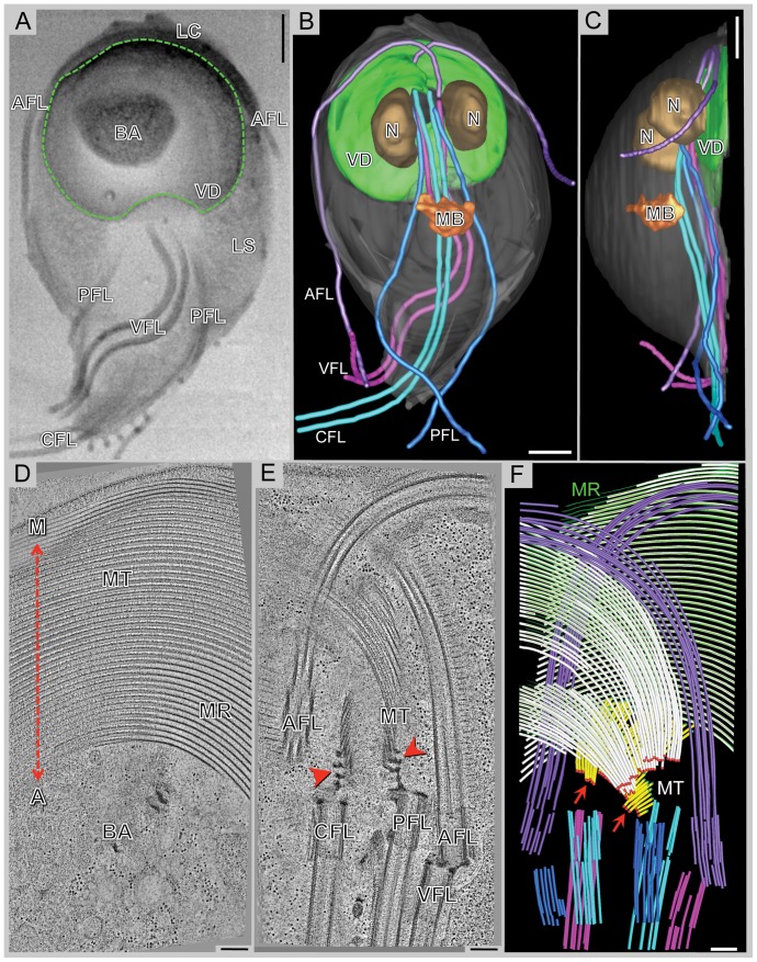
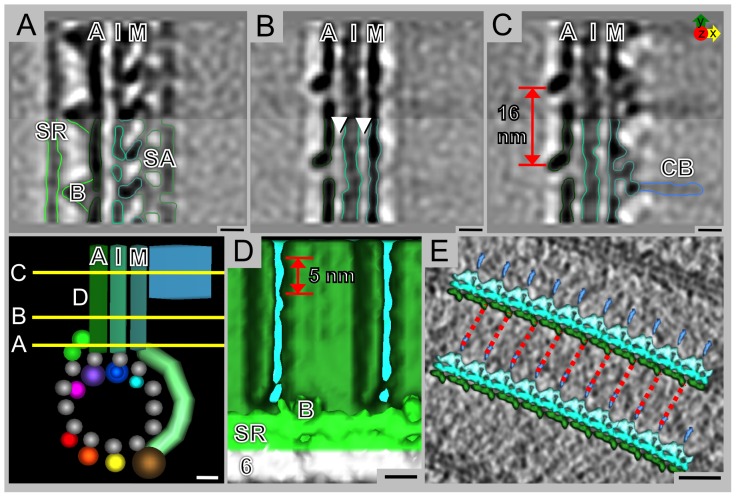
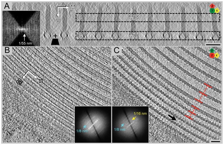
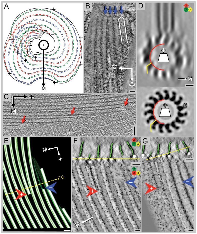
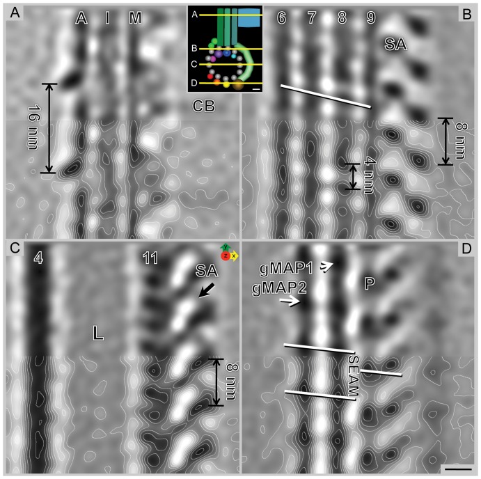
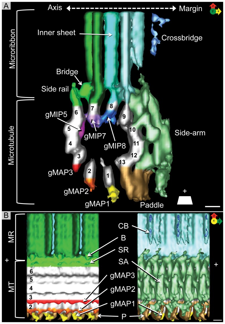
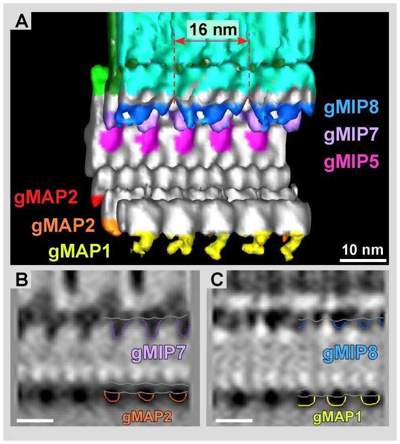
Giardia lamblia is a flagellated, unicellular parasite of mammals infecting over one billion people worldwide. Giardia's two-stage life cycle includes a motile trophozoite stage that colonizes the host small intestine and an infectious cyst form that can persist in the environment. Similar to many eukaryotic cells, Giardia contains several complex microtubule arrays that are involved in motility, chromosome segregation, organelle transport, maintenance of cell shape and transformation between the two life cycle stages. Giardia trophozoites also possess a unique spiral microtubule array, the ventral disc, made of approximately 50 parallel microtubules and associated microribbons, as well as a variety of associated proteins. The ventral disc maintains trophozoite attachment to the host intestinal epithelium. With the help of a combined SEM/microtome based slice and view method called 3View® (Gatan Inc., Pleasanton, CA), we present an entire trophozoite cell reconstruction and describe the arrangement of the major cytoskeletal elements. To aid in future analyses of disc-mediated attachment, we used electron-tomography of freeze-substituted, plastic-embedded trophozoites to explore the detailed architecture of ventral disc microtubules and their associated components. Lastly, we examined the disc microtubule array in three dimensions in unprecedented detail using cryo-electron tomography combined with internal sub-tomogram volume averaging of repetitive domains. We discovered details of protein complexes stabilizing microtubules by attachment to their inner and outer wall. A unique tri-laminar microribbon structure is attached vertically to the disc microtubules and is connected to neighboring microribbons via crossbridges. This work provides novel insight into the structure of the ventral disc microtubules, microribbons and associated proteins. Knowledge of the components comprising these structures and their three-dimensional organization is crucial toward understanding how attachment via the ventral disc occurs in vivo.
蓝氏贾第鞭毛虫是一种有鞭毛的、单细胞的哺乳动物寄生虫,感染了全球超过 10 亿人。贾第虫的两阶段生命周期包括一个运动的营养体阶段,它定殖在宿主的小肠,和一个传染性的囊形成,它可以在环境中持续存在。与许多真核细胞一样,贾第虫含有几个复杂的微管阵列,这些微管阵列参与运动、染色体分离、细胞器运输、细胞形状的维持和两个生命周期阶段之间的转化。贾第虫营养体还拥有一个独特的螺旋微管阵列,腹盘,由大约 50 个平行的微管和相关的微丝以及各种相关的蛋白质组成。腹盘维持营养体附着在宿主的肠上皮上。借助一种称为 3View®(Gatan Inc.,Pleasanton,CA)的基于 SEM/切片机的切片和视图组合方法,我们呈现了一个完整的营养体细胞重建,并描述了主要细胞骨架元件的排列。为了帮助未来对盘介导的附着进行分析,我们使用冷冻取代、塑料包埋的营养体的电子断层扫描来探索腹盘微管及其相关成分的详细结构。最后,我们使用冷冻电子断层扫描结合内部亚断层体积平均重复结构域,以前所未有的细节检查了盘微管的三维排列。我们发现了通过附着在微管的内、外壁上稳定微管的蛋白质复合物的细节。一个独特的三层微丝结构垂直附着在盘微管上,并通过交联桥与相邻的微丝相连。这项工作提供了关于腹盘微管、微丝和相关蛋白结构的新见解。了解构成这些结构的成分及其三维组织对于理解活体中通过腹盘附着的发生机制至关重要。