• 文献检索
  • 文档翻译
  • 深度研究
  • 学术资讯
  • Suppr Zotero 插件Zotero 插件
  • 邀请有礼
  • 套餐&价格
  • 历史记录
应用&插件
Suppr Zotero 插件Zotero 插件浏览器插件Mac 客户端Windows 客户端微信小程序
定价
高级版会员购买积分包购买API积分包
服务
文献检索文档翻译深度研究API 文档MCP 服务
关于我们
关于 Suppr公司介绍联系我们用户协议隐私条款
关注我们

Suppr 超能文献

核心技术专利:CN118964589B侵权必究
粤ICP备2023148730 号-1Suppr @ 2026

文献检索

告别复杂PubMed语法,用中文像聊天一样搜索,搜遍4000万医学文献。AI智能推荐,让科研检索更轻松。

立即免费搜索

文件翻译

保留排版,准确专业,支持PDF/Word/PPT等文件格式,支持 12+语言互译。

免费翻译文档

深度研究

AI帮你快速写综述,25分钟生成高质量综述,智能提取关键信息,辅助科研写作。

立即免费体验

儿童脑裂头蚴病:流行病学、临床和磁共振成像特征。

Cerebral sparganosis in children: epidemiological, clinical and MR imaging characteristics.

机构信息

Department of Radiology, Xiangya hospital, Central South University, Changsha, Hunan, 410008, China.

出版信息

BMC Pediatr. 2012 Sep 24;12:155. doi: 10.1186/1471-2431-12-155.

DOI:10.1186/1471-2431-12-155
PMID:23006504
原文链接:https://pmc.ncbi.nlm.nih.gov/articles/PMC3484034/
Abstract

BACKGROUND

Cerebral sparganosis in children is an extremely rare disease of central nervous system, and caused by a tapeworm larva from the genus of Spirometra. In this study, we discussed and summarized epidemiological, clinical and MR imaging characteristics of eighteen children with cerebral sparganosis for a better diagnosis and treatment of the disease.

METHODS

Eighteen children with cerebral sparganosis verified by pathology, serological tests and MR presentations were retrospectively investigated, and the epidemiologic and clinical characteristics of the disease were studied.

RESULTS

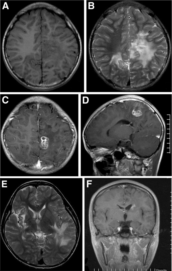
Twenty-seven lesions were found in the eighteen children. Twelve lesions in twelve patients were solitary while the lesions in the rest six patients were multiple and asymmetrical. The positions of the lesions were: seven in frontal, eleven in parietal, four in temporal and two in occipital lobes, one in basal ganglia, one in cerebella hemisphere and one in pons. The lesions were presented as slight hypointensity on T1-weighted images but moderate hyperintensity on T2-weighted images with perilesional brain parenchyma edema. Enhanced MR scans by using Gadopentetic Acid Dimeglumine Salt were performed in the patients, and the images demonstrated abnormal enhancements with the patterns of a peripheral ring, or a tortuous beaded, or a serpiginous tubular shape. Follow-up MR scans were preformed for eight patients, and three out of the eight cases exposed migrations and changes in shapes of the lesion areas.

CONCLUSIONS

The MR presentations in our study in general were similar to those in previous studies. However serpiginous tubular and comma-shaped enhancements of lesions have not been previously reported. The enhanced MR imaging and follow-up MR scans with the positive results from serological tests are the most important methods for the clinical diagnosis of cerebral sparganosis in children.

摘要

背景

儿童脑裂头蚴病是一种极为罕见的中枢神经系统寄生虫病,由裂头蚴属的绦虫幼虫引起。本研究通过讨论和总结 18 例儿童脑裂头蚴病的流行病学、临床和磁共振成像(MRI)特征,旨在提高对该病的诊断和治疗水平。

方法

回顾性分析 18 例经病理、血清学检查和 MRI 证实的脑裂头蚴病患儿的临床资料,总结其流行病学和临床特征。

结果

18 例患儿共发现 27 个病灶。12 例患儿的 12 个病灶为单发,其余 6 例患儿的病灶为多发且不对称,病变部位:额叶 7 个,顶叶 11 个,颞叶 4 个,枕叶 2 个,基底节区 1 个,小脑半球 1 个,脑桥 1 个。病灶 T1WI 呈稍低信号,T2WI 呈等或稍高信号,周围脑实质水肿。增强 MRI 扫描均采用钆喷酸葡胺,病灶呈异常强化,强化形式有:周边环状、迂曲串珠状或蜿蜒管状。8 例患儿进行了随访 MRI 扫描,其中 3 例病灶部位出现迁移和形态改变。

结论

本研究中患儿的 MRI 表现与既往研究基本相似,但未见呈蜿蜒管状和逗号形强化的病灶。增强 MRI 成像和随访 MRI 扫描,结合血清学检查阳性结果,是儿童脑裂头蚴病的重要临床诊断方法。

https://cdn.ncbi.nlm.nih.gov/pmc/blobs/f5d6/3484034/f9bb15f7c6c0/1471-2431-12-155-6.jpg
https://cdn.ncbi.nlm.nih.gov/pmc/blobs/f5d6/3484034/ff47d2e90d8a/1471-2431-12-155-1.jpg
https://cdn.ncbi.nlm.nih.gov/pmc/blobs/f5d6/3484034/e3291a445c48/1471-2431-12-155-2.jpg
https://cdn.ncbi.nlm.nih.gov/pmc/blobs/f5d6/3484034/fb0b28c59388/1471-2431-12-155-3.jpg
https://cdn.ncbi.nlm.nih.gov/pmc/blobs/f5d6/3484034/bc9f93a5685a/1471-2431-12-155-4.jpg
https://cdn.ncbi.nlm.nih.gov/pmc/blobs/f5d6/3484034/a09e4e268b09/1471-2431-12-155-5.jpg
https://cdn.ncbi.nlm.nih.gov/pmc/blobs/f5d6/3484034/f9bb15f7c6c0/1471-2431-12-155-6.jpg

相似文献

1
Cerebral sparganosis in children: epidemiological, clinical and MR imaging characteristics.儿童脑裂头蚴病:流行病学、临床和磁共振成像特征。
BMC Pediatr. 2012 Sep 24;12:155. doi: 10.1186/1471-2431-12-155.
2
[MRI diagnosis of cerebral sparganosis in children].[儿童脑裂头蚴病的磁共振成像诊断]
Zhongguo Dang Dai Er Ke Za Zhi. 2008 Aug;10(4):481-4.
3
MR spectroscopy and MR perfusion character of cerebral sparganosis: a case report.脑裂头蚴病的磁共振波谱和磁共振灌注特征:一例报告。
Br J Radiol. 2010 Feb;83(986):e31-4. doi: 10.1259/bjr/73038348.
4
Cerebral sparganosis: MR imaging versus CT features.脑裂头蚴病:磁共振成像与CT表现对比
Radiology. 1993 Sep;188(3):751-7. doi: 10.1148/radiology.188.3.8351344.
5
Migration: a notable feature of cerebral sparganosis on follow-up MR imaging.迁移:脑裂头蚴病随访磁共振成像的一个显著特征。
AJNR Am J Neuroradiol. 2013 Feb;34(2):327-33. doi: 10.3174/ajnr.A3237. Epub 2012 Aug 2.
6
Magnetic resonance imaging features of pathologically proven cerebral sparganosis.经病理证实的脑裂头蚴病的磁共振成像特征
J Int Med Res. 2013 Jun;41(3):867-77. doi: 10.1177/0300060513480925. Epub 2013 May 16.
7
CT and MR characteristics of cerebral sparganosis.脑裂头蚴病的CT及磁共振成像特征
AJNR Am J Neuroradiol. 2007 Oct;28(9):1700-5. doi: 10.3174/ajnr.A0659. Epub 2007 Sep 20.
8
Cerebral Sparganosis in Children: Epidemiologic and Radiologic Characteristics and Treatment Outcomes: A Report of 9 Cases.儿童脑裂头蚴病:流行病学、放射学特征及治疗结果:9例报告
World Neurosurg. 2016 May;89:153-8. doi: 10.1016/j.wneu.2016.01.086. Epub 2016 Feb 6.
9
[Cerebral sparganosis in a child: case report].[儿童脑裂头蚴病:病例报告]
Arch Argent Pediatr. 2013 Jan-Feb;111(1):e1-4. doi: 10.5546/aap.2013.e1.
10
Letter to the editor: role of imaging in the diagnosis of cerebral sparganosis.致编辑的信:影像学在脑裂头蚴病诊断中的作用
Br J Radiol. 2011 May;84(1001):481. doi: 10.1259/bjr/13076379.

引用本文的文献

1
Recent update on cerebral sparganosis: A bibliometric analysis and scientific mapping.脑裂头蚴病的最新进展:文献计量分析与科学图谱
Narra J. 2024 Aug;4(2):e982. doi: 10.52225/narra.v4i2.982. Epub 2024 Aug 15.
2
Cerebral Sparganosis - An Unusual Parasitic Infection Mimicking Cerebral Tuberculosis: Isolation of a Live Plerocercoid Larva of Spirometra mansoni.脑裂头蚴病——一种酷似脑结核的罕见寄生虫感染:曼氏迭宫绦虫活裂头蚴的分离
Ann Indian Acad Neurol. 2024 Jul 1;27(4):443-447. doi: 10.4103/aian.aian_2_24. Epub 2024 Apr 17.
3
Surgical treatment of a patient with live intracranial sparganosis for 17 years.

本文引用的文献

1
Letter to the editor: role of imaging in the diagnosis of cerebral sparganosis.致编辑的信:影像学在脑裂头蚴病诊断中的作用
Br J Radiol. 2011 May;84(1001):481. doi: 10.1259/bjr/13076379.
2
Diagnosis and stereotactic aspiration treatment of cerebral sparganosis: summary of 11 cases.脑裂头蚴病的诊断和立体定向抽吸治疗:11 例总结。
J Neurosurg. 2011 May;114(5):1421-5. doi: 10.3171/2010.4.JNS1079.
3
MR spectroscopy and MR perfusion character of cerebral sparganosis: a case report.脑裂头蚴病的磁共振波谱和磁共振灌注特征:一例报告。
17 年颅内裂头蚴存活患者的外科治疗。
BMC Infect Dis. 2022 Apr 9;22(1):353. doi: 10.1186/s12879-022-07293-7.
4
Anaplastic pleomorphic xanthoastrocytoma misdiagnosed as cerebral sparganosis-identification of the "mirror image".误诊为脑裂头蚴病的间变性多形性黄色星形细胞瘤——“镜像”的识别
Quant Imaging Med Surg. 2021 Oct;11(10):4479-4487. doi: 10.21037/qims-20-1398.
5
A Retrieved Sparganum of Spirometra erinaceieuropaei from a Korean Man during Mechanical Thrombectomy.一名韩国男性在机械取栓过程中取出的曼氏迭宫绦虫裂头蚴
Korean J Parasitol. 2020 Jun;58(3):309-313. doi: 10.3347/kjp.2020.58.3.309. Epub 2020 Jun 26.
6
Occurrence of Kalicephalus, Strongyloides, and Rhabdias nematodes as most common gastrointestinal parasites in captive snakes of German households and zoological gardens.在德国家庭和动物园饲养的蛇类中,Kalicephalus、Strongyloides 和 Rhabdias 线虫是最常见的胃肠道寄生虫。
Parasitol Res. 2020 Mar;119(3):947-956. doi: 10.1007/s00436-019-06526-0. Epub 2020 Jan 16.
7
Follow-up study of high-dose praziquantel therapy for cerebral sparganosis.大剂量吡喹酮治疗脑囊虫病的随访研究。
PLoS Negl Trop Dis. 2019 Jan 14;13(1):e0007018. doi: 10.1371/journal.pntd.0007018. eCollection 2019 Jan.
8
Occurrence of health-compromising protozoan and helminth infections in tortoises kept as pet animals in Germany.在德国,作为宠物饲养的龟类中存在危害健康的原生动物和蠕虫感染。
Parasit Vectors. 2018 Jun 18;11(1):352. doi: 10.1186/s13071-018-2936-z.
9
Orbital sparganosis in an 8-year boy: a case report.一名8岁男孩的眼眶裂头蚴病:病例报告
BMC Ophthalmol. 2018 Jan 22;18(1):13. doi: 10.1186/s12886-018-0675-8.
10
A waterborn zoonotic helminthiase in an Italian diver: a case report of a cutaneous Sparganum infection and a review of European cases.一名意大利潜水员感染的水源性人畜共患蠕虫病:一例皮肤曼氏裂头蚴感染病例报告及欧洲病例综述
Pathog Glob Health. 2015;109(8):383-6. doi: 10.1080/20477724.2015.1123901. Epub 2016 Jan 9.
Br J Radiol. 2010 Feb;83(986):e31-4. doi: 10.1259/bjr/73038348.
4
Cerebral sparganosis: the wandering lesion.脑裂头蚴病:游走性病变
Neurology. 2010 Jan 12;74(2):180. doi: 10.1212/WNL.0b013e3181c91a15.
5
CT and MR characteristics of cerebral sparganosis.脑裂头蚴病的CT及磁共振成像特征
AJNR Am J Neuroradiol. 2007 Oct;28(9):1700-5. doi: 10.3174/ajnr.A0659. Epub 2007 Sep 20.
6
Neuroimaging and pathological findings in a child with cerebral sparganosis. Case report.一名脑裂头蚴病患儿的神经影像学和病理学表现。病例报告。
J Neurosurg. 2006 Dec;105(6 Suppl):470-2. doi: 10.3171/ped.2006.105.6.470.
7
A review of human sparganosis in Thailand.泰国人体裂头蚴病综述。
Int J Infect Dis. 2005 Nov;9(6):312-6. doi: 10.1016/j.ijid.2004.08.003. Epub 2005 Jul 14.
8
Sparganosis in male breast.
J Formos Med Assoc. 2005 Feb;104(2):127-8.
9
Serological Diagnosis Of Human Sparganosis By Means Of Micro-ELISA.应用微量酶联免疫吸附试验进行人体裂头蚴病的血清学诊断
Kisaengchunghak Chapchi. 1984 Dec;22(2):222-228. doi: 10.3347/kjp.1984.22.2.222.
10
Cerebral sparganosis in a child.一名儿童的脑裂头蚴病。
Pediatr Neurosurg. 1997 Feb;26(2):103-6. doi: 10.1159/000121171.