Kato Takashi, Yamaguchi Koji, Kinoshita Koji, Sasaki Kiyotaka, Kagaya Hidetoshi, Meguro Takashi, Morita Takayuki, Takahashi Toshiyuki, Tamaki Nagara, Horita Shoichi
Department of Internal Medicine, Hokkaido Gastroenterology Hospital, Sapporo, Japan.
Case Rep Gastroenterol. 2012 Sep;6(3):754-9. doi: 10.1159/000346285. Epub 2012 Dec 19.
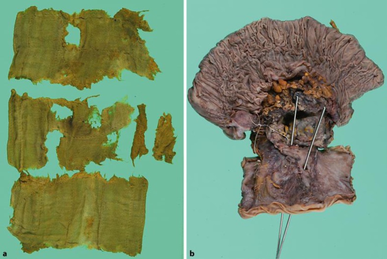
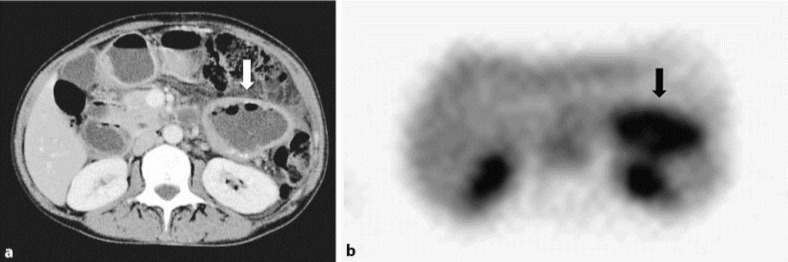
A 56-year-old woman with a history of gynecological surgery for cervical cancer 18 years previously was referred to our hospital for colicky abdominal pain, nausea and vomiting. Intestinal obstruction was diagnosed by contrast-enhanced computed tomography (CT) which showed dilation of the small intestine and suggested obstruction in the terminal ileum. In addition, CT showed a thick-walled cavitary lesion communicating with the proximal jejunum. (18)F-fluorodeoxyglucose positron emission tomography showed abnormal uptake at the same location as the cavitary lesion revealed by CT. The patient underwent laparotomy for the ileus and resection of the cavitary lesion. At laparotomy, we found a retained surgical sponge in the ileum 60 cm from the ileocecal valve. The cavitary tumor had two fistulae communicating with the proximal jejunum. The tumor was resected en bloc together with the transverse colon, part of the jejunum and the duodenum. Microscopic examination revealed fibrous encapsulation and foreign body giant cell reaction. Since a retained surgical sponge without radiopaque markers is extremely difficult to diagnose, retained surgical sponge should be considered in the differential diagnosis of intestinal obstruction in patients who have undergone previous abdominal surgery.
一名56岁女性,18年前因宫颈癌接受过妇科手术,因腹部绞痛、恶心和呕吐被转诊至我院。通过对比增强计算机断层扫描(CT)诊断为肠梗阻,CT显示小肠扩张,并提示回肠末端梗阻。此外,CT显示一个厚壁空洞性病变与空肠近端相通。(18)F-氟脱氧葡萄糖正电子发射断层扫描显示,病变部位的摄取情况与CT显示的空洞性病变位置相同。患者因肠梗阻接受剖腹手术,并切除了空洞性病变。剖腹手术时,我们在距回盲瓣60厘米的回肠中发现了一块残留的手术海绵。空洞性肿瘤有两个与空肠近端相通的瘘管。肿瘤与横结肠、部分空肠和十二指肠一起整块切除。显微镜检查显示有纤维包裹和异物巨细胞反应。由于没有不透射线标记物的残留手术海绵极难诊断,对于既往有腹部手术史的肠梗阻患者,鉴别诊断时应考虑残留手术海绵的可能。