Department of Neuroscience, University of Pittsburgh Pittsburgh, PA, USA.
Front Neurosci. 2013 Jan 21;6:199. doi: 10.3389/fnins.2012.00199. eCollection 2012.
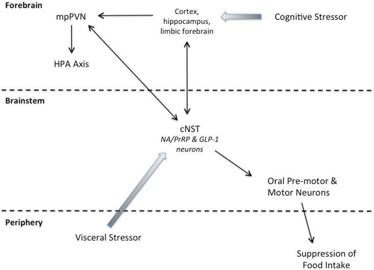
Neural circuits distributed within the brainstem, hypothalamus, and limbic forebrain interact to control food intake and energy balance under normal day-to-day conditions, and in response to stressful conditions under which homeostasis is threatened. Experimental studies using rats and mice have generated a voluminous literature regarding the functional organization of circuits that inhibit food intake in response to satiety signals, and in response to stress. Although the central neural bases of satiation and stress-induced hypophagia often are studied and discussed as if they were distinct, we propose that both behavioral states are generated, at least in part, by recruitment of two separate but intermingled groups of caudal hindbrain neurons. One group comprises a subpopulation of noradrenergic (NA) neurons within the caudal nucleus of the solitary tract (cNST; A2 cell group) that is immunopositive for prolactin-releasing peptide (PrRP). The second group comprises non-adrenergic neurons within the cNST and nearby reticular formation that synthesize glucagon-like peptide 1 (GLP-1). Axonal projections from PrRP and GLP-1 neurons target distributed brainstem and forebrain regions that shape behavioral, autonomic, and endocrine responses to actual or anticipated homeostatic challenge, including the challenge of food intake. Evidence reviewed in this article supports the view that hindbrain PrRP and GLP-1 neurons contribute importantly to satiation and stress-induced hypophagia by modulating the activity of caudal brainstem circuits that control food intake. Hindbrain PrRP and GLP-1 neurons also engage hypothalamic and limbic forebrain networks that drive parallel behavioral and endocrine functions related to food intake and homeostatic challenge, and modulate conditioned and motivational aspects of food intake.
分布在脑干、下丘脑和边缘前脑的神经网络相互作用,在正常的日常条件下以及在威胁内稳态的应激条件下,控制食物摄入和能量平衡。使用大鼠和小鼠的实验研究产生了大量关于抑制食物摄入的回路的功能组织的文献,这些回路对饱腹感信号和应激做出反应。尽管饱食和应激诱导的摄食减少的中枢神经基础通常被研究和讨论为截然不同的状态,但我们提出,这两种行为状态至少部分是由两组独立但交织的尾后脑神经元募集产生的。一组包括尾侧孤束核(cNST;A2 细胞群)内的去甲肾上腺素能(NA)神经元亚群,该神经元对催乳素释放肽(PrRP)呈免疫阳性。第二组包括 cNST 内和附近的网状结构中的非肾上腺素能神经元,它们合成胰高血糖素样肽 1(GLP-1)。PrRP 和 GLP-1 神经元的轴突投射靶向分布在脑干和前脑的区域,这些区域塑造了对实际或预期的稳态挑战(包括食物摄入的挑战)的行为、自主和内分泌反应。本文综述的证据支持这样一种观点,即尾侧脑 PrRP 和 GLP-1 神经元通过调节控制食物摄入的尾侧脑干回路的活动,对饱食和应激诱导的摄食减少做出重要贡献。尾侧脑 PrRP 和 GLP-1 神经元还参与驱动与食物摄入和稳态挑战相关的平行行为和内分泌功能的下丘脑和边缘前脑网络,并调节条件和动机方面的食物摄入。