Laboratorio de Fisiología y Plasticidad Neuronal, Departamento de Fisiología, Facultad de Biología, Universidad de Sevilla, Sevilla, Spain.
PLoS One. 2013;8(1):e54519. doi: 10.1371/journal.pone.0054519. Epub 2013 Jan 18.
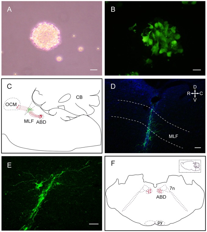
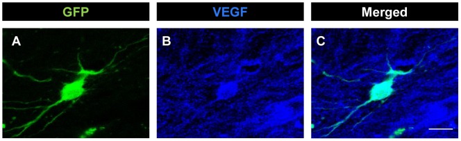
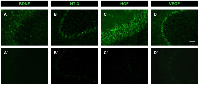
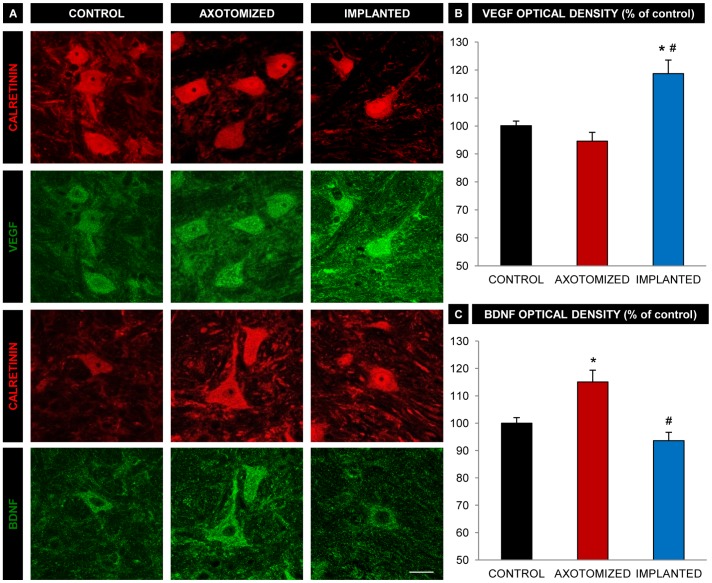
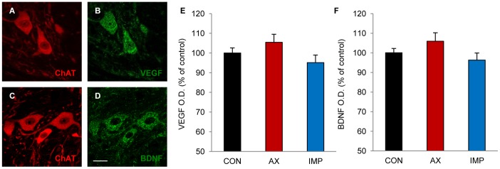
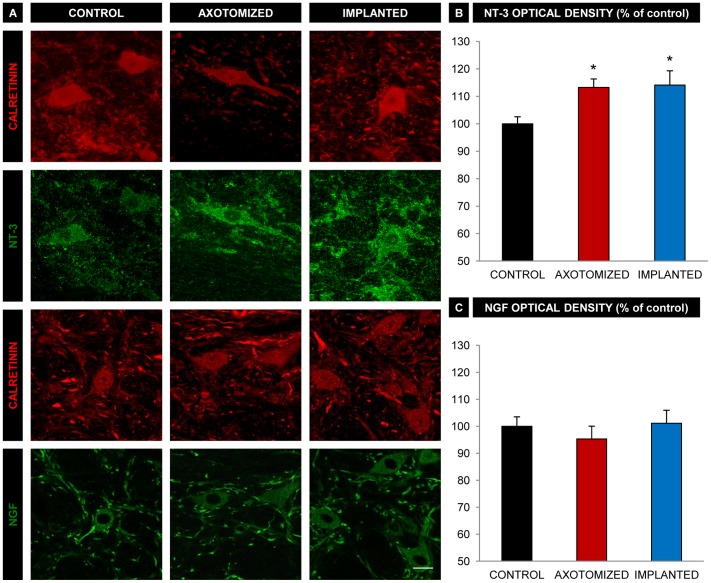
Axotomy of central neurons leads to functional and structural alterations which largely revert when neural progenitor cells (NPCs) are implanted in the lesion site. The new microenvironment created by NPCs in the host tissue might modulate in the damaged neurons the expression of a high variety of molecules with relevant roles in the repair mechanisms, including neurotrophic factors. In the present work, we aimed to analyze changes in neurotrophic factor expression in axotomized neurons induced by NPC implants. For this purpose, we performed immunofluorescence followed by confocal microscopy analysis for the detection of vascular endothelial growth factor (VEGF), brain-derived neurotrophic factor (BDNF), neurotrophin-3 (NT-3) and nerve growth factor (NGF) on brainstem sections from rats with axotomy of abducens internuclear neurons that received NPC implants (implanted group) or vehicle injections (axotomized group) in the lesion site. Control abducens internuclear neurons were strongly immunoreactive to VEGF and BDNF but showed a weak staining for NT-3 and NGF. Comparisons between groups revealed that lesioned neurons from animals that received NPC implants showed a significant increase in VEGF content with respect to animals receiving vehicle injections. However, the immunoreactivity for BDNF, which was increased in the axotomized group as compared to control, was not modified in the implanted group. The modifications induced by NPC implants on VEGF and BDNF content were specific for the population of axotomized abducens internuclear neurons since the neighboring abducens motoneurons were not affected. Similar levels of NT-3 and NGF immunolabeling were obtained in injured neurons from axotomized and implanted animals. Among all the analyzed neurotrophic factors, only VEGF was expressed by the implanted cells in the lesion site. Our results point to a role of NPC implants in the modulation of neurotrophic factor expression by lesioned central neurons, which might contribute to the restorative effects of these implants.
中枢神经元的轴突切断会导致功能和结构的改变,而当神经祖细胞(NPC)被植入损伤部位时,这些改变会在很大程度上恢复。NPC 在宿主组织中形成的新微环境可能会调节损伤神经元中表达的大量与修复机制相关的分子,包括神经营养因子。在本工作中,我们旨在分析 NPC 植入诱导的轴突切断神经元中神经营养因子表达的变化。为此,我们进行了免疫荧光 followed by 共聚焦显微镜分析,以检测脑桥切片中血管内皮生长因子(VEGF)、脑源性神经营养因子(BDNF)、神经营养因子-3(NT-3)和神经生长因子(NGF)的表达,这些切片来自接受 NPC 植入(植入组)或损伤部位注射载体(切断组)的外展核间神经元切断的大鼠。对照外展核间神经元对 VEGF 和 BDNF 具有强烈的免疫反应性,但对 NT-3 和 NGF 的染色较弱。组间比较显示,接受 NPC 植入的动物的损伤神经元中 VEGF 含量显著增加,而接受载体注射的动物则没有。然而,与对照相比,在切断组中增加的 BDNF 免疫反应性在植入组中没有改变。NPC 植入对 VEGF 和 BDNF 含量的影响是特定于切断的外展核间神经元群体的,因为相邻的外展运动神经元没有受到影响。在切断和植入动物的损伤神经元中,获得了相似水平的 NT-3 和 NGF 免疫标记。在分析的所有神经营养因子中,只有 VEGF 是由损伤部位的植入细胞表达的。我们的结果表明 NPC 植入在调节损伤中枢神经元神经营养因子表达方面发挥作用,这可能有助于这些植入物的修复效果。