Astrahan Melvin A
Department of Radiation Oncology, University of Southern California School of Medicine, Los Angeles and Eye Physics, LLC, Los Alamitos, California, USA.
J Contemp Brachytherapy. 2013 Mar;5(1):23-32. doi: 10.5114/jcb.2013.34450. Epub 2013 Mar 29.
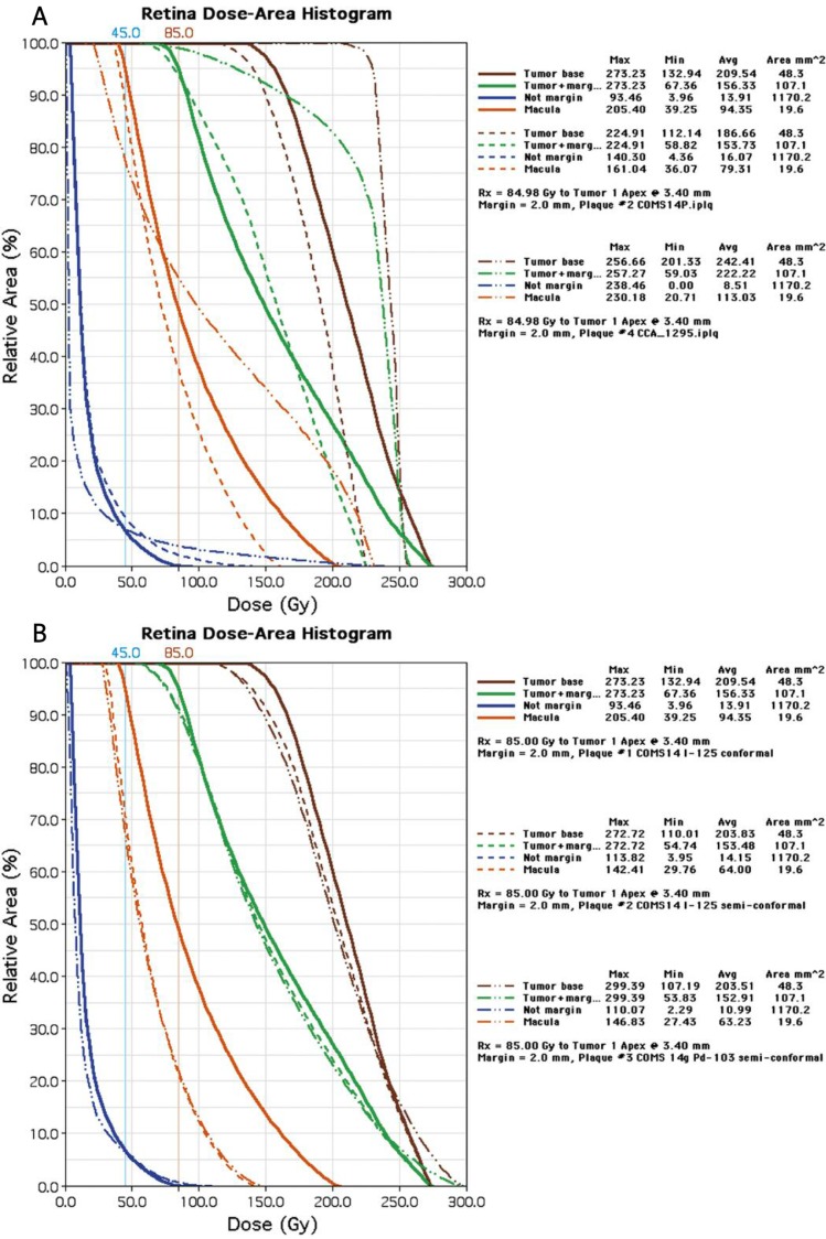
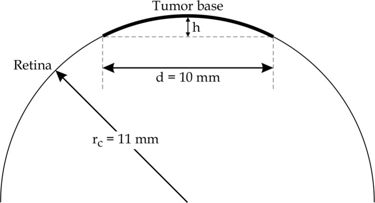
Episcleral plaques have a history of over a half century in the delivery of radiation therapy to intraocular tumors such as choroidal melanoma. Although the tumor control rate is high, vision-impairing complications subsequent to treatment remain an issue. Notable, late complications are radiation retinopathy and maculopathy. The obvious way to reduce the risk of radiation damage to the retina is to conform the prescribed isodose surface to the tumor base and to reduce the dose delivered to the surrounding healthy retina, especially the macula. Using a fusion of fundus photography, ultrasound and CT images, tumor size, shape and location within the eye can be accurately simulated as part of the radiation planning process. In this work an adaptation of the dose-volume histogram (DVH), the retina dose-area histogram (RDAH) is introduced as a metric to help compare rival plaque designs and conformal treatment planning options with the goal of reducing radiation retinopathy.
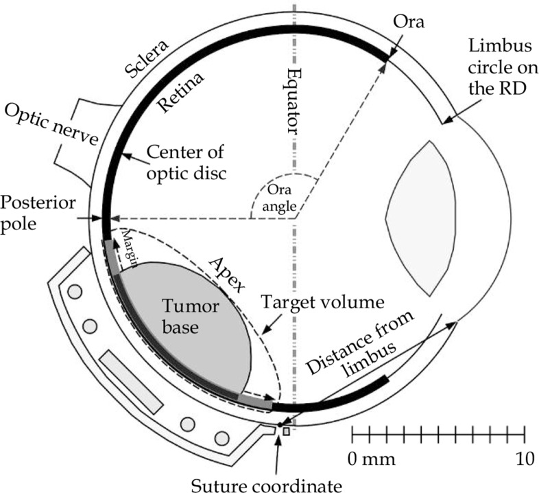
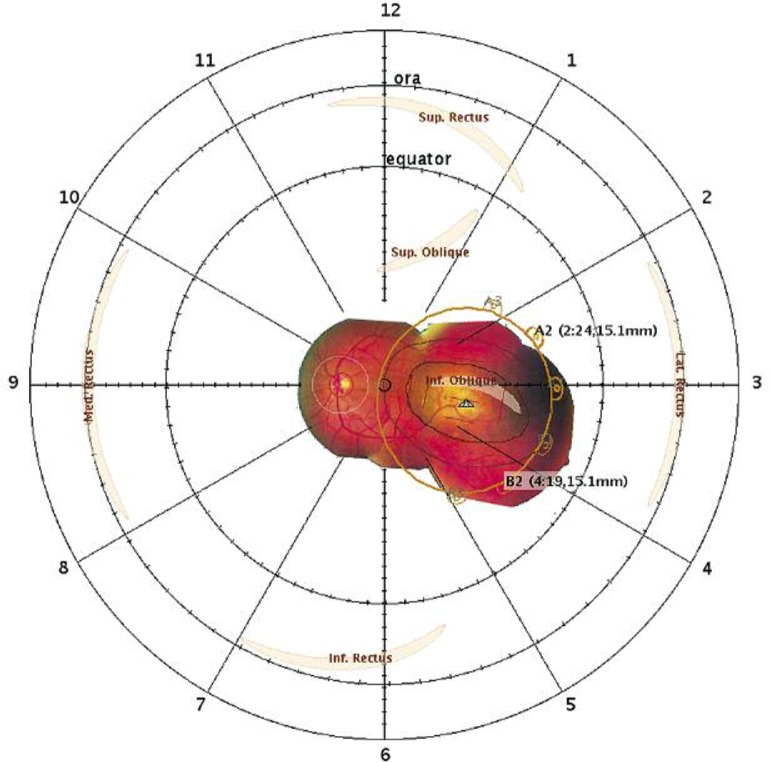
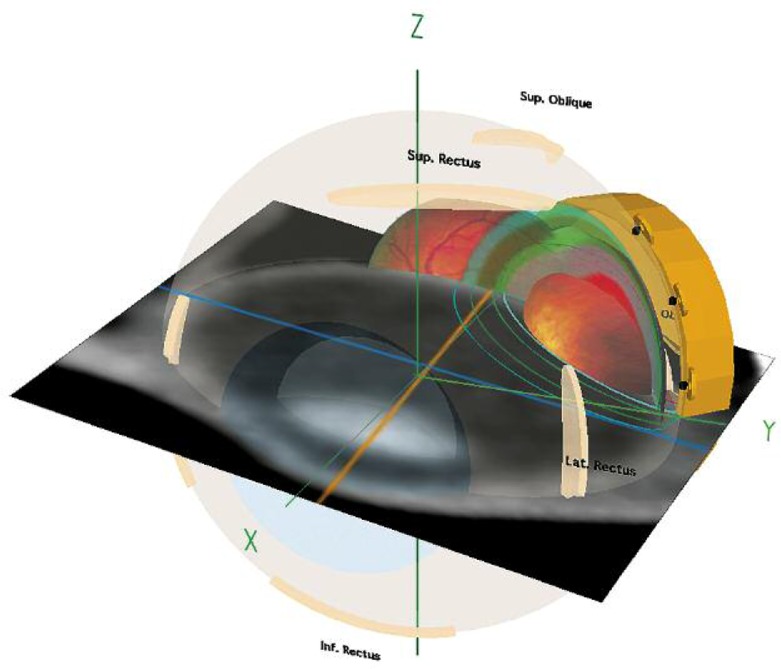
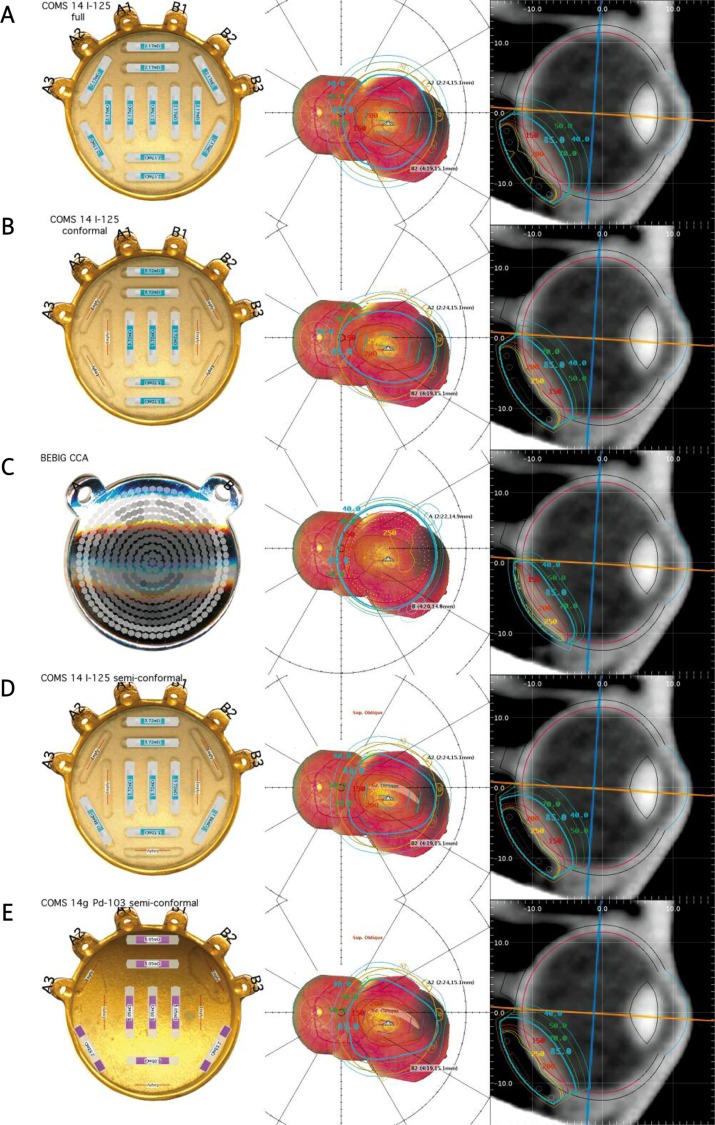
The RDAH is calculated by transforming a digitized fundus-photo collage of the tumor into a rasterized polar map of the retinal surface known as a retinal diagram (RD). The perimeter of the tumor base is digitized on the RD and its area computed. Area and radiation dose are calculated for every pixel in the RD.
The areal resolution of the RDAH is a function of the pixel resolution of the raster image used to display the RD and the number of polygon edges used to digitize the perimeter of the tumor base. A practical demonstration is presented.
The RDAH provides a quantitative metric by which episcleral plaque treatment plan options may be evaluated and compared in order to confirm adequate dosimetric coverage of the tumor and margin, and to help minimize dose to the macula and retina.
巩膜外斑块用于脉络膜黑色素瘤等眼内肿瘤的放射治疗已有半个多世纪的历史。尽管肿瘤控制率很高,但治疗后导致视力受损的并发症仍然是一个问题。值得注意的是,晚期并发症是放射性视网膜病变和黄斑病变。降低视网膜辐射损伤风险的明显方法是使规定的等剂量表面与肿瘤基底相符,并减少传递到周围健康视网膜,尤其是黄斑的剂量。通过融合眼底摄影、超声和CT图像,可以在放射治疗计划过程中准确模拟眼内肿瘤的大小、形状和位置。在这项工作中,引入了剂量体积直方图(DVH)的一种变体——视网膜剂量面积直方图(RDAH)作为一种度量标准,以帮助比较不同的斑块设计和适形治疗计划方案,目标是减少放射性视网膜病变。
RDAH通过将肿瘤的数字化眼底照片拼贴转换为视网膜表面的光栅化极坐标图(称为视网膜图,RD)来计算。在RD上对肿瘤基底的周长进行数字化处理并计算其面积。计算RD中每个像素的面积和辐射剂量。
RDAH的面积分辨率取决于用于显示RD的光栅图像的像素分辨率以及用于数字化肿瘤基底周长的多边形边数。给出了一个实际示例。
RDAH提供了一种定量度量标准,通过它可以评估和比较巩膜外斑块治疗计划方案,以确认肿瘤及其边缘有足够的剂量覆盖,并有助于将黄斑和视网膜的剂量降至最低。