• 文献检索
  • 文档翻译
  • 深度研究
  • 学术资讯
  • Suppr Zotero 插件Zotero 插件
  • 邀请有礼
  • 套餐&价格
  • 历史记录
应用&插件
Suppr Zotero 插件Zotero 插件浏览器插件Mac 客户端Windows 客户端微信小程序
定价
高级版会员购买积分包购买API积分包
服务
文献检索文档翻译深度研究API 文档MCP 服务
关于我们
关于 Suppr公司介绍联系我们用户协议隐私条款
关注我们

Suppr 超能文献

核心技术专利:CN118964589B侵权必究
粤ICP备2023148730 号-1Suppr @ 2026

文献检索

告别复杂PubMed语法,用中文像聊天一样搜索,搜遍4000万医学文献。AI智能推荐,让科研检索更轻松。

立即免费搜索

文件翻译

保留排版,准确专业,支持PDF/Word/PPT等文件格式,支持 12+语言互译。

免费翻译文档

深度研究

AI帮你快速写综述,25分钟生成高质量综述,智能提取关键信息,辅助科研写作。

立即免费体验

人角膜内皮细胞培养的优化:体外成功培养的密度依赖性

Optimization of human corneal endothelial cell culture: density dependency of successful cultures in vitro.

作者信息

Peh Gary S L, Toh Kah-Peng, Ang Heng-Pei, Seah Xin-Yi, George Benjamin L, Mehta Jodhbir S

机构信息

Tissue Engineering and Stem Cell Group, Singapore Eye Research Institute, 11 Third Hospital Ave, #06-00, Singapore 168751, Singapore.

出版信息

BMC Res Notes. 2013 May 3;6:176. doi: 10.1186/1756-0500-6-176.

DOI:10.1186/1756-0500-6-176
PMID:23641909
原文链接:https://pmc.ncbi.nlm.nih.gov/articles/PMC3659058/
Abstract

BACKGROUND

Global shortage of donor corneas greatly restricts the numbers of corneal transplantations performed yearly. Limited ex vivo expansion of primary human corneal endothelial cells is possible, and a considerable clinical interest exists for development of tissue-engineered constructs using cultivated corneal endothelial cells. The objective of this study was to investigate the density-dependent growth of human corneal endothelial cells isolated from paired donor corneas and to elucidate an optimal seeding density for their extended expansion in vitro whilst maintaining their unique cellular morphology.

RESULTS

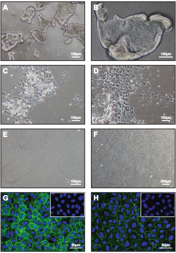
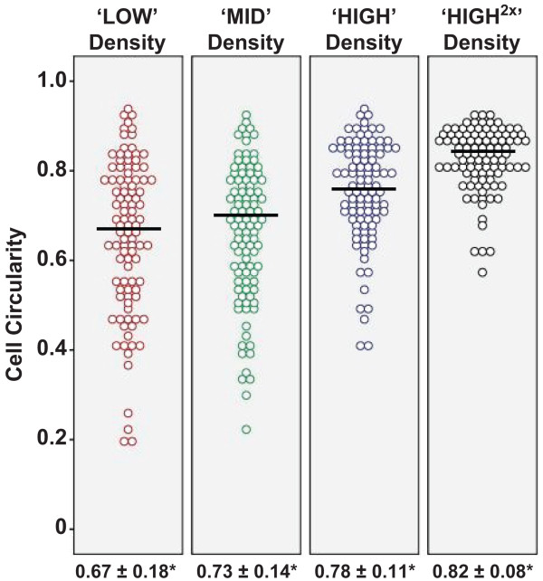
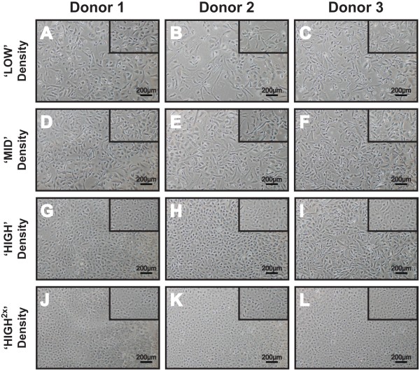
Established primary human corneal endothelial cells were propagated to the second passage (P2) before they were utilized for this study. Confluent P2 cells were dissociated and seeded at four seeding densities: 2,500 cells per cm2 ('LOW'); 5,000 cells per cm2 ('MID'); 10,000 cells per cm2 ('HIGH'); and 20,000 cells per cm2 ('HIGH(×2)'), and subsequently analyzed for their propensity to proliferate. They were also subjected to morphometric analyses comparing cell sizes, coefficient of variance, as well as cell circularity when each culture became confluent. At the two lower densities, proliferation rates were higher than cells seeded at higher densities, though not statistically significant. However, corneal endothelial cells seeded at lower densities were significantly larger in size, heterogeneous in shape and less circular (fibroblastic-like), and remained hypertrophic after one month in culture. Comparatively, cells seeded at higher densities were significantly homogeneous, compact and circular at confluence. Potentially, at an optimal seeding density of 10,000 cells per cm2, it is possible to obtain between 10 million to 25 million cells at the third passage. More importantly, these expanded human corneal endothelial cells retained their unique cellular morphology.

CONCLUSIONS

Our results demonstrated a density dependency in the culture of primary human corneal endothelial cells. Sub-optimal seeding density results in a decrease in cell saturation density, as well as a loss in their proliferative potential. As such, we propose a seeding density of not less than 10,000 cells per cm2 for regular passage of primary human corneal endothelial cells.

摘要

背景

供体角膜的全球短缺极大地限制了每年进行的角膜移植数量。原代人角膜内皮细胞的体外扩增有限,因此利用培养的角膜内皮细胞开发组织工程构建体具有相当大的临床意义。本研究的目的是研究从配对供体角膜分离的人角膜内皮细胞的密度依赖性生长,并阐明其在体外扩展培养时保持独特细胞形态的最佳接种密度。

结果

已建立的原代人角膜内皮细胞在用于本研究之前传代至第二代(P2)。将汇合的P2细胞解离并以四种接种密度接种:每平方厘米2500个细胞(“低”);每平方厘米5000个细胞(“中”);每平方厘米10000个细胞(“高”);以及每平方厘米20000个细胞(“高(×2)”),随后分析它们的增殖倾向。当每种培养物汇合时,还对它们进行形态计量分析,比较细胞大小、变异系数以及细胞圆形度。在两个较低密度下,增殖率高于以较高密度接种的细胞,尽管无统计学意义。然而,以较低密度接种的角膜内皮细胞尺寸明显更大,形状异质且圆形度较低(成纤维细胞样),并且在培养一个月后仍呈肥大状态。相比之下,以较高密度接种的细胞在汇合时明显均匀、紧密且呈圆形。潜在地,在每平方厘米10000个细胞的最佳接种密度下,第三代时有可能获得1000万至2500万个细胞。更重要的是,这些扩增的人角膜内皮细胞保留了其独特的细胞形态。

结论

我们的结果证明了原代人角膜内皮细胞培养中的密度依赖性。接种密度次优会导致细胞饱和密度降低以及增殖潜能丧失。因此,我们建议原代人角膜内皮细胞常规传代的接种密度不低于每平方厘米10000个细胞。

https://cdn.ncbi.nlm.nih.gov/pmc/blobs/6a72/3659058/daa23ea74fc9/1756-0500-6-176-3.jpg
https://cdn.ncbi.nlm.nih.gov/pmc/blobs/6a72/3659058/bc1ebf6def14/1756-0500-6-176-1.jpg
https://cdn.ncbi.nlm.nih.gov/pmc/blobs/6a72/3659058/6f5a941aa307/1756-0500-6-176-2.jpg
https://cdn.ncbi.nlm.nih.gov/pmc/blobs/6a72/3659058/daa23ea74fc9/1756-0500-6-176-3.jpg
https://cdn.ncbi.nlm.nih.gov/pmc/blobs/6a72/3659058/bc1ebf6def14/1756-0500-6-176-1.jpg
https://cdn.ncbi.nlm.nih.gov/pmc/blobs/6a72/3659058/6f5a941aa307/1756-0500-6-176-2.jpg
https://cdn.ncbi.nlm.nih.gov/pmc/blobs/6a72/3659058/daa23ea74fc9/1756-0500-6-176-3.jpg

相似文献

1
Optimization of human corneal endothelial cell culture: density dependency of successful cultures in vitro.人角膜内皮细胞培养的优化:体外成功培养的密度依赖性
BMC Res Notes. 2013 May 3;6:176. doi: 10.1186/1756-0500-6-176.
2
Corneal Endothelial Cell Cultures from Organotypic Preservation of Older Donor Corneas Are Suitable for Advanced Cell Therapy.从老年供体角膜的器官型保存中培养的角膜内皮细胞适合用于高级细胞疗法。
Ophthalmic Res. 2023;66(1):1254-1265. doi: 10.1159/000533701. Epub 2023 Sep 18.
3
Cultivation of human corneal endothelial cells isolated from paired donor corneas.从配对供体角膜中分离培养人角膜内皮细胞。
PLoS One. 2011;6(12):e28310. doi: 10.1371/journal.pone.0028310. Epub 2011 Dec 16.
4
[Transplantation of corneal endothelial cells].[角膜内皮细胞移植]
Nippon Ganka Gakkai Zasshi. 2002 Dec;106(12):805-35; discussion 836.
5
Properties of corneas reconstructed with cultured human corneal endothelial cells and human corneal stroma.用人角膜内皮细胞和人角膜基质重建的角膜的特性。
Jpn J Ophthalmol. 2005 Nov-Dec;49(6):448-452. doi: 10.1007/s10384-005-0245-5.
6
Passaging capability of human corneal endothelial cells derived from old donors with and without accelerating cell attachment.具有和不具有加速细胞附着能力的老年供体来源的人角膜内皮细胞的传代能力。
Exp Eye Res. 2019 Dec;189:107814. doi: 10.1016/j.exer.2019.107814. Epub 2019 Sep 24.
7
Propagation of human corneal endothelial cells: a novel dual media approach.人角膜内皮细胞的增殖:一种新型双培养基方法。
Cell Transplant. 2015;24(2):287-304. doi: 10.3727/096368913X675719. Epub 2013 Nov 21.
8
Transplantation of adult human or porcine corneal endothelial cells onto human recipients in vitro. Part I: Cell culturing and transplantation procedure.将成人人类或猪角膜内皮细胞体外移植到人类受体。第一部分:细胞培养与移植程序。
Cornea. 1999 Mar;18(2):199-206. doi: 10.1097/00003226-199903000-00010.
9
Improving the success rate of human corneal endothelial cell cultures from single donor corneas with stabilization medium.使用稳定培养基提高单供体角膜人角膜内皮细胞培养的成功率。
Cell Tissue Bank. 2018 Mar;19(1):9-17. doi: 10.1007/s10561-017-9665-y. Epub 2017 Oct 17.
10
Impact of culture media on primary human corneal endothelial cells derived from old donors.培养基于老供体的原代人角膜内皮细胞的培养液影响因素
Exp Eye Res. 2024 Mar;240:109815. doi: 10.1016/j.exer.2024.109815. Epub 2024 Feb 3.

引用本文的文献

1
Preclinical Models for Studying Fuchs Endothelial Corneal Dystrophy.用于研究富克斯角膜内皮营养不良的临床前模型
Cells. 2025 Mar 28;14(7):505. doi: 10.3390/cells14070505.
2
An effective method for culturing functional human corneal endothelial cells using a xenogeneic free culture medium.一种使用无异种培养物的培养基培养功能性人眼角膜内皮细胞的有效方法。
Sci Rep. 2023 Nov 9;13(1):19492. doi: 10.1038/s41598-023-46590-2.
3
Biomaterials used for tissue engineering of barrier-forming cell monolayers in the eye.用于眼部屏障形成细胞单层组织工程的生物材料。

本文引用的文献

1
NIH Image to ImageJ: 25 years of image analysis.NIH 图像到 ImageJ:25 年的图像分析。
Nat Methods. 2012 Jul;9(7):671-5. doi: 10.1038/nmeth.2089.
2
ROCK inhibitor converts corneal endothelial cells into a phenotype capable of regenerating in vivo endothelial tissue.ROCK 抑制剂将角膜内皮细胞转化为具有体内再生内皮组织能力的表型。
Am J Pathol. 2012 Jul;181(1):268-77. doi: 10.1016/j.ajpath.2012.03.033. Epub 2012 Jun 14.
3
Cultivation of human corneal endothelial cells isolated from paired donor corneas.从配对供体角膜中分离培养人角膜内皮细胞。
Front Bioeng Biotechnol. 2023 Sep 27;11:1269385. doi: 10.3389/fbioe.2023.1269385. eCollection 2023.
4
Investigating the Role of TGF-β Signaling Pathways in Human Corneal Endothelial Cell Primary Culture.研究 TGF-β 信号通路在人眼角膜内皮细胞原代培养中的作用。
Cells. 2023 Jun 14;12(12):1624. doi: 10.3390/cells12121624.
5
Ocular-Surface Regeneration Therapies for Eye Disorders: The State of the Art.眼部疾病的眼表再生疗法:现状
BioTech (Basel). 2023 Jun 15;12(2):48. doi: 10.3390/biotech12020048.
6
Effects of Rho-Associated Kinase (Rock) Inhibitors (Alternative to Y-27632) on Primary Human Corneal Endothelial Cells.Rho 相关激酶(Rock)抑制剂(替代 Y-27632)对原代人角膜内皮细胞的影响。
Cells. 2023 May 3;12(9):1307. doi: 10.3390/cells12091307.
7
Isolation efficiency of collagenase and EDTA for the culture of corneal endothelial cells.胶原酶和 EDTA 对角膜内皮细胞培养的分离效率。
Mol Vis. 2022 Oct 2;28:331-339. eCollection 2022.
8
Ex vivo expansion and characterization of human corneal endothelium for transplantation: a review.人眼角膜内皮细胞的离体扩增与鉴定用于移植:综述。
Stem Cell Res Ther. 2021 Oct 30;12(1):554. doi: 10.1186/s13287-021-02611-3.
9
Challenges in corneal endothelial cell culture.角膜内皮细胞培养的挑战。
Regen Med. 2021 Sep;16(9):871-891. doi: 10.2217/rme-2020-0202. Epub 2021 Aug 12.
10
Experimental design of a culture approach for corneal endothelial cells of New Zealand white rabbit.新西兰白兔角膜内皮细胞培养方法的实验设计
Heliyon. 2020 Oct 6;6(10):e05178. doi: 10.1016/j.heliyon.2020.e05178. eCollection 2020 Oct.
PLoS One. 2011;6(12):e28310. doi: 10.1371/journal.pone.0028310. Epub 2011 Dec 16.
4
Development of new therapeutic modalities for corneal endothelial disease focused on the proliferation of corneal endothelial cells using animal models.针对角膜内皮细胞增殖的角膜内皮疾病的新治疗方法的开发,使用动物模型。
Exp Eye Res. 2012 Feb;95(1):60-7. doi: 10.1016/j.exer.2011.10.014. Epub 2011 Nov 3.
5
Proliferative capacity of corneal endothelial cells.角膜内皮细胞的增殖能力。
Exp Eye Res. 2012 Feb;95(1):16-23. doi: 10.1016/j.exer.2011.08.014. Epub 2011 Aug 30.
6
Human corneal endothelial cell expansion for corneal endothelium transplantation: an overview.人眼角膜内皮细胞的扩增用于角膜内皮移植:概述。
Transplantation. 2011 Apr 27;91(8):811-9. doi: 10.1097/TP.0b013e3182111f01.
7
Bioengineering endothelialized neo-corneas using donor-derived corneal endothelial cells and decellularized corneal stroma.利用供体来源的角膜内皮细胞和去细胞化的角膜基质生物工程化内皮neo 角膜。
Biomaterials. 2010 Sep;31(26):6738-45. doi: 10.1016/j.biomaterials.2010.05.020. Epub 2010 Jun 11.
8
Quantifying pharmacologic suppression of cellular senescence: prevention of cellular hypertrophy versus preservation of proliferative potential.量化细胞衰老的药理学抑制:预防细胞肥大与保留增殖潜能。
Aging (Albany NY). 2009 Dec 31;1(12):1008-16. doi: 10.18632/aging.100115.
9
Assessment of attachment factors for primary cultured human corneal endothelial cells.评估原代培养人角膜内皮细胞的附着因子。
Cornea. 2009 Oct;28(9):1050-4. doi: 10.1097/ICO.0b013e3181a165a3.
10
Enhancement on primate corneal endothelial cell survival in vitro by a ROCK inhibitor.ROCK抑制剂对灵长类动物角膜内皮细胞体外存活的增强作用。
Invest Ophthalmol Vis Sci. 2009 Aug;50(8):3680-7. doi: 10.1167/iovs.08-2634. Epub 2009 Apr 22.