Du Tao, Zou Xiangyu, Cheng Jun, Wu Shuai, Zhong Liang, Ju Guanqun, Zhu Jiang, Liu Guohua, Zhu Yingjian, Xia Shujie
Stem Cell Res Ther. 2013 Jun 4;4(3):59. doi: 10.1186/scrt215.
Based on some well-documented reports, we attempted to clarify the antifibrotic mechanisms of human Wharton's-jelly-derived mesenchymal stromal cells (WJ-MSCs) from the perspective of induction of hepatocyte growth factor (HGF) expression in tubular epithelial cells (TECs).
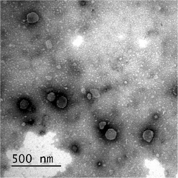
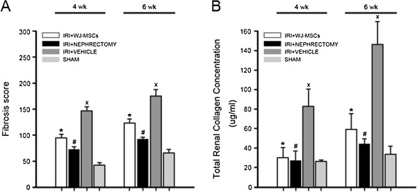
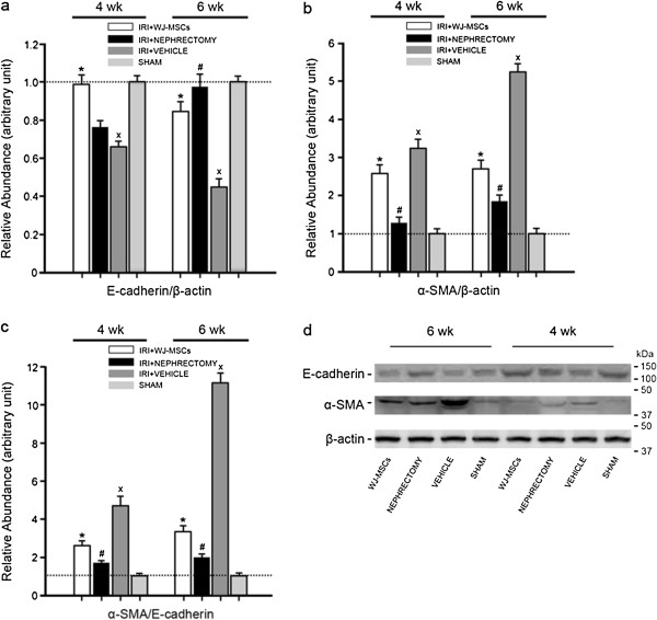
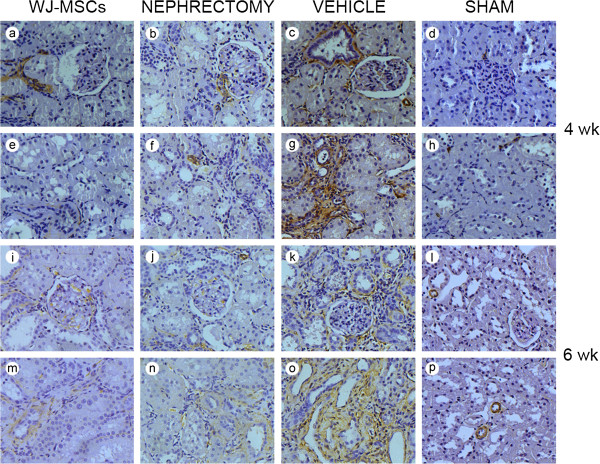
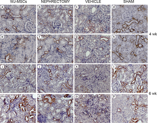
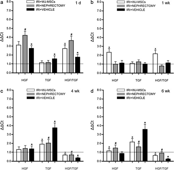
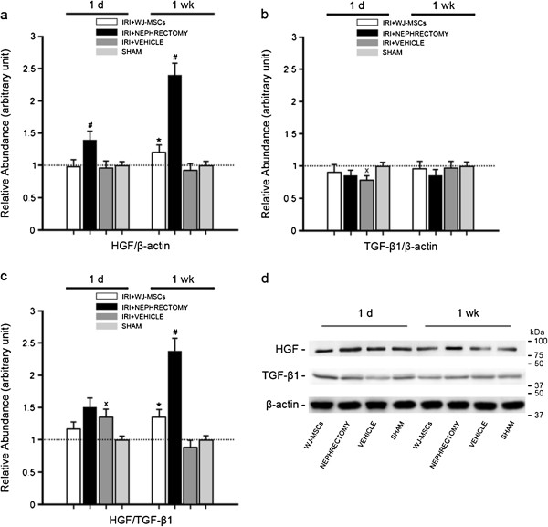
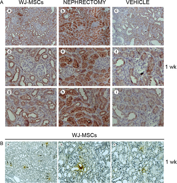
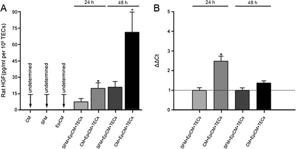
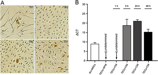
A rat model of acute kidney injury (AKI) was established through unilateral renal ischemia for 1 hour. Two days later, a single intravenous cell or vehicle injection, or contralateral nephrectomy, was performed. Rats were sacrificed at 1 day, 1 week, 4 weeks, or 6 weeks after the intervention. Renal fibrosis was evaluated by Masson trichrome staining and Sircol collagen assay. The upregulation of α-smooth muscle actin (α-SMA) versus E-cadherin expression was adopted as an indicator of tubular epithelial-mesenchymal transition (EMT). Gene and protein expression of HGF or transforming growth factor-beta1 (TGF-β1) was determined by real-time polymerase chain reaction (RT-PCR) and Western blot, respectively. HGF expression in TECs was detected with immunostaining. In vitro, rat TECs subjected to hypoxia injury were incubated with or without conditioned medium (CM) from WJ-MSCs for 1, 3, 24, or 48 hours. Rat or human HGF synthesis in TECs was assessed with immunostaining, RT-PCR, or ELISA.
Cell delivery or nephrectomy led to abrogation of renal scarring. At the incipient period of AKI, through induction of HGF expression, either of them remarkably promoted the upregulation of HGF versus TGF-β1 expression in damaged kidney. Rat TECs were not only the principal cells expressing HGF but also exhibited human HGF expression after cell infusion. During fibrogenesis, the downregulation of HGF versus TGF-β1 expression was greatly prevented by WJ-MSCs or kidney removal, thereby resulting in tubular EMT delay. In vitro, after 24 or 48 hours of incubation, CM not only robustly induced the upregulation of rat HGF gene expression in TECs but substantially amplified the release of rat HGF. Under the induction of CM, human HGF mRNA and protein were detected in rat TECs.
WJ-MSCs contribute to tubular EMT delay and the alleviation of renal fibrosis. Induction of native and foreign HGF synthesis in damaged TECs at the initial stage of AKI leads to recovery of the disturbed balance of HGF/TGF-β1 during scar formation, being one of the vital mechanisms.
基于一些有充分文献记载的报告,我们试图从诱导肾小管上皮细胞(TECs)中肝细胞生长因子(HGF)表达的角度,阐明人脐带华通氏胶间充质基质细胞(WJ-MSCs)的抗纤维化机制。
通过单侧肾脏缺血1小时建立大鼠急性肾损伤(AKI)模型。两天后,进行单次静脉注射细胞或注射溶媒,或对侧肾切除术。在干预后1天、1周、4周或6周处死大鼠。通过Masson三色染色和Sircol胶原测定法评估肾纤维化。采用α-平滑肌肌动蛋白(α-SMA)与E-钙黏蛋白表达的上调作为肾小管上皮-间充质转化(EMT)的指标。分别通过实时聚合酶链反应(RT-PCR)和蛋白质印迹法测定HGF或转化生长因子-β1(TGF-β1)的基因和蛋白表达。用免疫染色检测TECs中HGF的表达。在体外,将遭受缺氧损伤的大鼠TECs与有或无WJ-MSCs条件培养基(CM)孵育1、3、24或48小时。用免疫染色、RT-PCR或酶联免疫吸附测定(ELISA)评估TECs中大鼠或人HGF的合成。
细胞输注或肾切除术可消除肾瘢痕形成。在AKI初期,通过诱导HGF表达,二者均显著促进受损肾脏中HGF相对于TGF-β1表达的上调。大鼠TECs不仅是表达HGF的主要细胞,而且在细胞输注后还表现出人HGF表达。在纤维化形成过程中,WJ-MSCs或肾切除极大地阻止了HGF相对于TGF-β1表达的下调,从而导致肾小管EMT延迟。在体外,孵育24或48小时后,CM不仅强烈诱导TECs中大鼠HGF基因表达上调,而且大幅增加大鼠HGF的释放。在CM诱导下,在大鼠TECs中检测到了人HGF mRNA和蛋白。
WJ-MSCs有助于延缓肾小管EMT并减轻肾纤维化。在AKI初期诱导受损TECs合成内源性和外源性HGF,可使瘢痕形成过程中HGF/TGF-β1失衡得以恢复,这是重要机制之一。