Centre of clinical investigation in biotherapy CIC-BT 507, Institut Gustave Roussy, Villejuif, France.
Br J Cancer. 2013 Aug 20;109(4):1013-22. doi: 10.1038/bjc.2013.362. Epub 2013 Jul 18.
The aim of our study was to evaluate the prognostic role of immunological microenvironnement in stage II-III CRC patients.
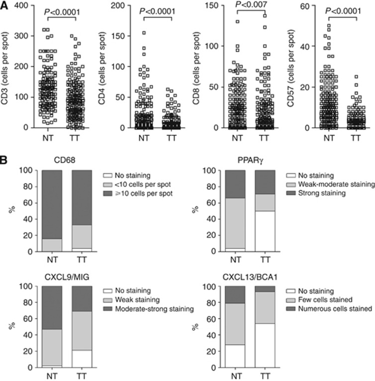
We constructed a tissue microarray from 196 consecutive patients with stage II-III CRC and compared CD3, CD4, CD8, CD57, CD68, CXCL9/MIG, CXCL13, and PPARγ immunoreactivity in tumour samples and their matched non-tumour tissue. We assessed their association with relapse-free survival (RFS; primary endpoint) and overall survival (OS) in multivariate Cox models.
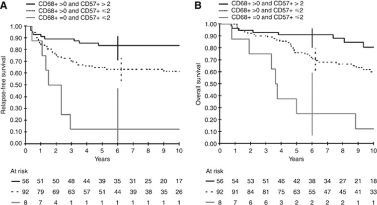
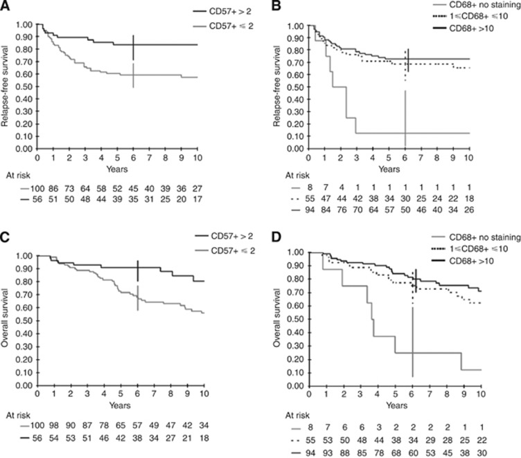
Low densities of CD57+ and CD68+ tumour-infiltrating cells (TIC) independently predicted worse outcomes. A prognostic score combining CD57 (+, > vs -, ≤2 cells per spot) and CD68 (+, >0 vs -, =0 cells per spot) TIC density discriminated CRC patients at low (CD68+/CD57+), intermediate (CD68+/CD57-), or high (CD68-/CD57-) risk, with hazard ratios for the intermediate-risk and high-risk groups of 2.7 (95% confidence interval (CI): 1.3-5.8) and 9.0 (3.2-25.4) for RFS, and 2.5 (1.2-5.1) and 10.6 (3.8-29.2) for OS, respectively, as compared with the low-risk group. Corresponding 5-year survival rates (95% CI) in the low-, moderate- and high-risk groups were 84% (71-91), 65% (54-74), and 12% (2-47), respectively, for RFS, and 91% (80-96), 76% (66-84), and 25% (7-59), respectively, for OS.
Tumour CD57+ and CD68+ TIC density assessment independently predicts survival in patients with stage II-III CRC. If validated, our score based on a quick, inexpensive, and well-established method such as point counting on diagnostic tissue sections could be used routinely as a prognostic tool in CRC patients.
本研究旨在评估免疫微环境在 II-III 期 CRC 患者中的预后作用。
我们从 196 例连续的 II-III 期 CRC 患者中构建了组织微阵列,并比较了肿瘤样本及其匹配的非肿瘤组织中 CD3、CD4、CD8、CD57、CD68、CXCL9/MIG、CXCL13 和 PPARγ 的免疫反应性。我们在多变量 Cox 模型中评估了它们与无复发生存率(RFS;主要终点)和总生存率(OS)的关系。
低密度的 CD57+和 CD68+肿瘤浸润细胞(TIC)独立预测预后不良。结合 CD57(+,> vs -,≤2 个细胞/点)和 CD68(+,>0 vs -,=0 个细胞/点)TIC 密度的预后评分可将 CRC 患者分为低(CD68+/CD57+)、中(CD68+/CD57-)或高(CD68-/CD57-)风险组,中风险和高风险组的 RFS 危险比分别为 2.7(95%置信区间(CI):1.3-5.8)和 9.0(3.2-25.4),OS 分别为 2.5(1.2-5.1)和 10.6(3.8-29.2),与低风险组相比。低、中、高风险组的 5 年生存率(95%CI)分别为 RFS 84%(71-91)、65%(54-74)和 12%(2-47),OS 91%(80-96)、76%(66-84)和 25%(7-59)。
肿瘤 CD57+和 CD68+TIC 密度评估独立预测 II-III 期 CRC 患者的生存。如果得到验证,我们的评分基于快速、廉价且成熟的方法,如在诊断组织切片上进行点计数,可以常规用作 CRC 患者的预后工具。