Mietens Andrea, Tasch Sabine, Stammler Angelika, Konrad Lutz, Feuerstacke Caroline, Middendorff Ralf
Institute of Anatomy and Cell Biology, Justus-Liebig-University Giessen, Giessen, Germany.
Department of Gynecology and Obstetrics, Justus-Liebig-University Giessen, Giessen, Germany.
PLoS One. 2014 Mar 24;9(3):e92603. doi: 10.1371/journal.pone.0092603. eCollection 2014.
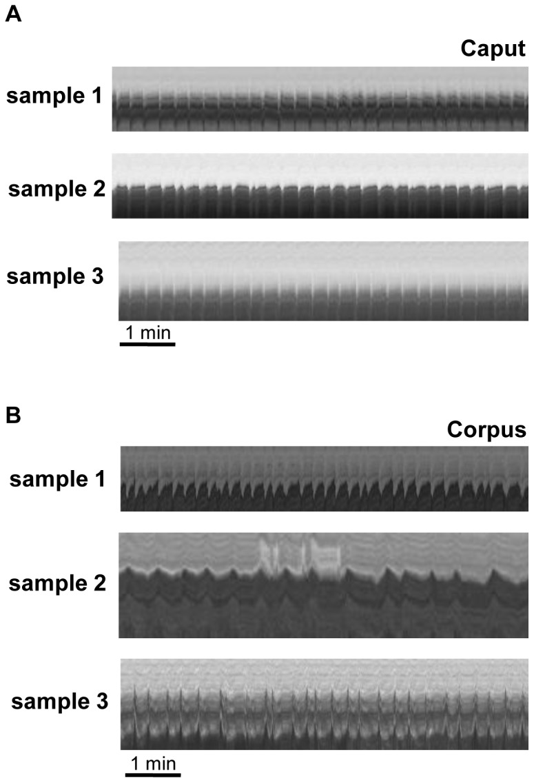
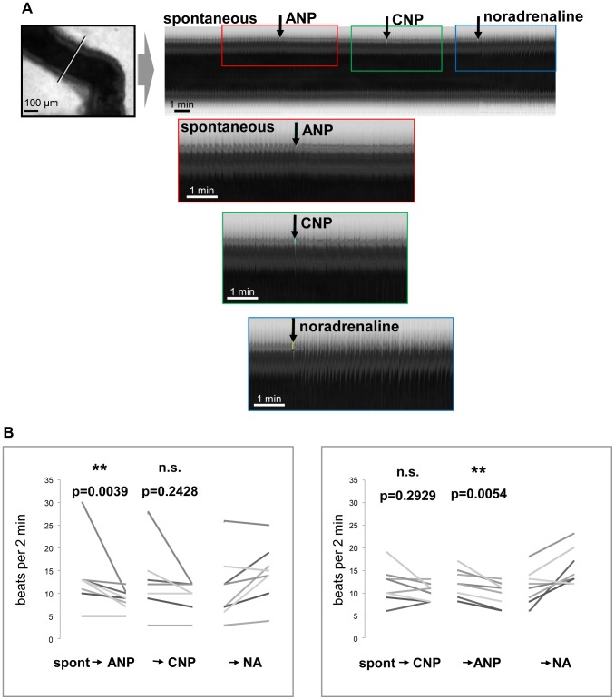
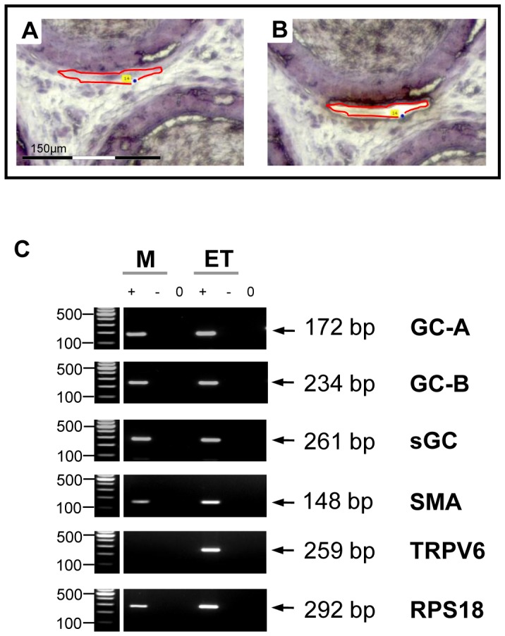
The well orchestrated function of epididymal smooth muscle cells ensures transit of spermatozoa through the epididymal duct during which spermatozoa acquire motility and fertilizing capacity. Relaxation of smooth muscle cells is mediated by cGMP signaling and components of this pathway are found within the male reproductive tract. Whereas contractile function of caudal parts of the rat epididymal duct can be examined in organ bath studies, caput and corpus regions are fragile and make it difficult to mount them in an organ bath. We developed an ex vivo time-lapse imaging-based approach to investigate the contractile pattern in these parts of the epididymal duct. Collagen-embedding allowed immobilization without impeding contractility or diffusion of drugs towards the duct and therefore facilitated subsequent movie analyses. The contractile pattern was made visible by placing virtual sections through the acquired image stack to track wall movements over time. By this, simultaneous evaluation of contractile activity at different positions of the observed duct segment was possible. With each contraction translating into a spike, drug-induced alterations in contraction frequency could be assessed easily. Peristaltic contractions were also detectable and throughout all regions in the proximal epididymis we found regular spontaneous contractile activity that elicited movement of intraluminal contents. Stimulating cGMP production by natriuretic peptide ANP or inhibiting degradation of cGMP by the phosphodiesterase 5 inhibitor sildenafil significantly reduced contractile frequency in isolated duct segments from caput and corpus. RT-PCR analysis after laser-capture microdissection localized the corresponding molecules to the smooth muscle layer of the duct. Our time-lapse imaging approach proved to be feasible to assess contractile function in all regions of the epididymal duct under near physiological conditions and provides a tool to evaluate acute (side) effects of drugs and to investigate various signaling pathways.
附睾平滑肌细胞精心编排的功能确保精子在附睾管中转运,在此过程中精子获得运动能力和受精能力。平滑肌细胞的舒张由cGMP信号介导,该信号通路的组成部分存在于男性生殖道内。虽然大鼠附睾管尾部的收缩功能可在器官浴实验中进行检测,但头部和体部区域较为脆弱,难以安装在器官浴中。我们开发了一种基于体外延时成像的方法来研究附睾管这些部位的收缩模式。胶原包埋可实现固定,同时不妨碍收缩性或药物向管道的扩散,因此便于后续的电影分析。通过在采集的图像堆栈中放置虚拟切片以跟踪壁随时间的运动,使收缩模式可见。通过这种方式,可以同时评估观察到的管道段不同位置的收缩活动。每次收缩转化为一个峰值,因此可以轻松评估药物引起的收缩频率变化。蠕动收缩也可检测到,在附睾近端的所有区域,我们都发现了规律的自发收缩活动,这种活动引起管腔内物质的移动。通过利钠肽ANP刺激cGMP生成或用磷酸二酯酶5抑制剂西地那非抑制cGMP降解,可显著降低来自头部和体部的离体管道段的收缩频率。激光捕获显微切割后的RT-PCR分析将相应分子定位到管道的平滑肌层。我们的延时成像方法被证明在接近生理条件下评估附睾管所有区域的收缩功能是可行的,并提供了一种评估药物急性(副作用)效应和研究各种信号通路的工具。