• 文献检索
  • 文档翻译
  • 深度研究
  • 学术资讯
  • Suppr Zotero 插件Zotero 插件
  • 邀请有礼
  • 套餐&价格
  • 历史记录
应用&插件
Suppr Zotero 插件Zotero 插件浏览器插件Mac 客户端Windows 客户端微信小程序
定价
高级版会员购买积分包购买API积分包
服务
文献检索文档翻译深度研究API 文档MCP 服务
关于我们
关于 Suppr公司介绍联系我们用户协议隐私条款
关注我们

Suppr 超能文献

核心技术专利:CN118964589B侵权必究
粤ICP备2023148730 号-1Suppr @ 2026

文献检索

告别复杂PubMed语法,用中文像聊天一样搜索,搜遍4000万医学文献。AI智能推荐,让科研检索更轻松。

立即免费搜索

文件翻译

保留排版,准确专业,支持PDF/Word/PPT等文件格式,支持 12+语言互译。

免费翻译文档

深度研究

AI帮你快速写综述,25分钟生成高质量综述,智能提取关键信息,辅助科研写作。

立即免费体验

蛋白激酶Cδ在血管内皮生长因子/趋化因子配体4诱导的内皮管解离过程中调节力信号传导。

PKCδ regulates force signaling during VEGF/CXCL4 induced dissociation of endothelial tubes.

作者信息

Jamison Joshua, Wang James H-C, Wells Alan

机构信息

Department of Pathology, McGowan Institute for Regenerative Medicine, University of Pittsburgh, Pittsburgh, Pennsylvania, United States of America.

Department of Orthopedic Surgery, McGowan Institute for Regenerative Medicine, University of Pittsburgh, Pittsburgh, Pennsylvania, United States of America.

出版信息

PLoS One. 2014 Apr 3;9(4):e93968. doi: 10.1371/journal.pone.0093968. eCollection 2014.

DOI:10.1371/journal.pone.0093968
PMID:24699667
原文链接:https://pmc.ncbi.nlm.nih.gov/articles/PMC3974837/
Abstract

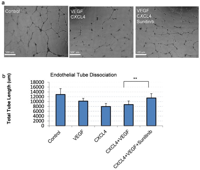
Wound healing requires the vasculature to re-establish itself from the severed ends; endothelial cells within capillaries must detach from neighboring cells before they can migrate into the nascent wound bed to initiate angiogenesis. The dissociation of these endothelial capillaries is driven partially by platelets' release of growth factors and cytokines, particularly the chemokine CXCL4/platelet factor-4 (PF4) that increases cell-cell de-adherence. As this retraction is partly mediated by increased transcellular contractility, the protein kinase c-δ/myosin light chain-2 (PKCδ/MLC-2) signaling axis becomes a candidate mechanism to drive endothelial dissociation. We hypothesize that PKCδ activation induces contractility through MLC-2 to promote dissociation of endothelial cords after exposure to platelet-released CXCL4 and VEGF. To investigate this mechanism of contractility, endothelial cells were allowed to form cords following CXCL4 addition to perpetuate cord dissociation. In this study, CXCL4-induced dissociation was reduced by a VEGFR inhibitor (sunitinib malate) and/or PKCδ inhibition. During combined CXCL4+VEGF treatment, increased contractility mediated by MLC-2 that is dependent on PKCδ regulation. As cellular force is transmitted to focal adhesions, zyxin, a focal adhesion protein that is mechano-responsive, was upregulated after PKCδ inhibition. This study suggests that growth factor regulation of PKCδ may be involved in CXCL4-mediated dissociation of endothelial cords.

摘要

伤口愈合需要血管系统从断端重新建立;毛细血管内的内皮细胞必须与相邻细胞分离,才能迁移到新生的伤口床以启动血管生成。这些内皮毛细血管的分离部分是由血小板释放的生长因子和细胞因子驱动的,特别是趋化因子CXCL4/血小板因子-4(PF4),它会增加细胞间的去黏附。由于这种收缩部分是由跨细胞收缩力增加介导的,蛋白激酶c-δ/肌球蛋白轻链-2(PKCδ/MLC-2)信号轴成为驱动内皮细胞分离的候选机制。我们假设PKCδ激活通过MLC-2诱导收缩,以促进内皮索在暴露于血小板释放的CXCL4和VEGF后分离。为了研究这种收缩机制,在添加CXCL4后让内皮细胞形成索,以延续索的分离。在本研究中,VEGFR抑制剂(苹果酸舒尼替尼)和/或PKCδ抑制可减少CXCL4诱导的分离。在联合CXCL4+VEGF处理期间,由MLC-2介导的收缩增加,这依赖于PKCδ调节。由于细胞力传递到黏着斑,黏着斑蛋白zyxin是机械反应性的,在PKCδ抑制后上调。这项研究表明,PKCδ的生长因子调节可能参与CXCL4介导的内皮索分离。

https://cdn.ncbi.nlm.nih.gov/pmc/blobs/3e53/3974837/2b54eea56146/pone.0093968.g005.jpg
https://cdn.ncbi.nlm.nih.gov/pmc/blobs/3e53/3974837/ec04987ad181/pone.0093968.g001.jpg
https://cdn.ncbi.nlm.nih.gov/pmc/blobs/3e53/3974837/dd700a4aa8de/pone.0093968.g002.jpg
https://cdn.ncbi.nlm.nih.gov/pmc/blobs/3e53/3974837/0299d21d906b/pone.0093968.g003.jpg
https://cdn.ncbi.nlm.nih.gov/pmc/blobs/3e53/3974837/05187c77753c/pone.0093968.g004.jpg
https://cdn.ncbi.nlm.nih.gov/pmc/blobs/3e53/3974837/2b54eea56146/pone.0093968.g005.jpg

相似文献

1
PKCδ regulates force signaling during VEGF/CXCL4 induced dissociation of endothelial tubes.蛋白激酶Cδ在血管内皮生长因子/趋化因子配体4诱导的内皮管解离过程中调节力信号传导。
PLoS One. 2014 Apr 3;9(4):e93968. doi: 10.1371/journal.pone.0093968. eCollection 2014.
2
Vascular endothelial growth factor (VEGF) and platelet (PF-4) factor 4 inputs modulate human microvascular endothelial signaling in a three-dimensional matrix migration context.血管内皮生长因子 (VEGF) 和血小板 (PF-4) 因子 4 输入调节人微血管内皮细胞在三维基质迁移环境中的信号转导。
Mol Cell Proteomics. 2013 Dec;12(12):3704-18. doi: 10.1074/mcp.M113.030528. Epub 2013 Sep 9.
3
PKCδ localization at the membrane increases matrix traction force dependent on PLCγ1/EGFR signaling.PKCδ 在膜上的定位增加了依赖于 PLCγ1/EGFR 信号的基质牵引力。
PLoS One. 2013 Oct 14;8(10):e77434. doi: 10.1371/journal.pone.0077434. eCollection 2013.
4
CXCL4L1 inhibits angiogenesis and induces undirected endothelial cell migration without affecting endothelial cell proliferation and monocyte recruitment.CXCL4L1 抑制血管生成,并诱导无定向的内皮细胞迁移,而不影响内皮细胞增殖和单核细胞募集。
J Thromb Haemost. 2011 Jan;9(1):209-19. doi: 10.1111/j.1538-7836.2010.04119.x.
5
Vascular endothelial growth factor-induced endothelial cell migration and proliferation depend on a nitric oxide-mediated decrease in protein kinase Cdelta activity.血管内皮生长因子诱导的内皮细胞迁移和增殖依赖于一氧化氮介导的蛋白激酶Cδ活性降低。
Circ Res. 1999 Aug 6;85(3):247-56. doi: 10.1161/01.res.85.3.247.
6
Thromboxane A2 receptor signaling inhibits vascular endothelial growth factor-induced endothelial cell differentiation and migration.血栓素A2受体信号传导抑制血管内皮生长因子诱导的内皮细胞分化和迁移。
Circ Res. 2004 Aug 20;95(4):372-9. doi: 10.1161/01.RES.0000138300.41642.15. Epub 2004 Jul 8.
7
Sunitinib inhibits lymphatic endothelial cell functions and lymph node metastasis in a breast cancer model through inhibition of vascular endothelial growth factor receptor 3.舒尼替尼通过抑制血管内皮生长因子受体 3 抑制乳腺癌模型中的淋巴管内皮细胞功能和淋巴结转移。
Breast Cancer Res. 2011 Jun 21;13(3):R66. doi: 10.1186/bcr2903.
8
Characterization of the metabolic changes underlying growth factor angiogenic activation: identification of new potential therapeutic targets.生长因子血管生成激活背后代谢变化的特征:新潜在治疗靶点的鉴定。
Carcinogenesis. 2009 Jun;30(6):946-52. doi: 10.1093/carcin/bgp083. Epub 2009 Apr 15.
9
Serum response factor is a critical requirement for VEGF signaling in endothelial cells and VEGF-induced angiogenesis.血清反应因子是内皮细胞中VEGF信号传导和VEGF诱导的血管生成的关键要素。
FASEB J. 2004 Aug;18(11):1264-6. doi: 10.1096/fj.03-1232fje. Epub 2004 Jun 4.
10
Protein kinase C-delta inhibition protects blood-brain barrier from sepsis-induced vascular damage.蛋白激酶 C-δ 抑制可保护血脑屏障免受脓毒症引起的血管损伤。
J Neuroinflammation. 2018 Nov 6;15(1):309. doi: 10.1186/s12974-018-1342-y.

引用本文的文献

1
In Vitro Comparison of Lymphangiogenic Potential of Hypoxia Preconditioned Serum (HPS) and Platelet-Rich Plasma (PRP).缺氧预处理血清(HPS)和富血小板血浆(PRP)的淋巴管生成潜力的体外比较。
Int J Mol Sci. 2023 Jan 19;24(3):1961. doi: 10.3390/ijms24031961.
2
Platelet proteome reveals novel pathways of platelet activation and platelet-mediated immunoregulation in dengue.血小板蛋白质组揭示了登革热中血小板活化及血小板介导的免疫调节的新途径。
PLoS Pathog. 2017 May 19;13(5):e1006385. doi: 10.1371/journal.ppat.1006385. eCollection 2017 May.
3
The Fibrin Matrix Regulates Angiogenic Responses within the Hemostatic Microenvironment through Biochemical Control.

本文引用的文献

1
PKCδ localization at the membrane increases matrix traction force dependent on PLCγ1/EGFR signaling.PKCδ 在膜上的定位增加了依赖于 PLCγ1/EGFR 信号的基质牵引力。
PLoS One. 2013 Oct 14;8(10):e77434. doi: 10.1371/journal.pone.0077434. eCollection 2013.
2
Interplay between FAK, PKCδ, and p190RhoGAP in the regulation of endothelial barrier function.FAK、PKCδ 和 p190RhoGAP 在调节内皮细胞屏障功能中的相互作用。
Microvasc Res. 2012 Jan;83(1):12-21. doi: 10.1016/j.mvr.2011.04.005. Epub 2011 Apr 22.
3
Pericyte actomyosin-mediated contraction at the cell-material interface can modulate the microvascular niche.
纤维蛋白基质通过生化控制调节止血微环境中的血管生成反应。
PLoS One. 2015 Aug 28;10(8):e0135618. doi: 10.1371/journal.pone.0135618. eCollection 2015.
4
Retinoblastoma binding protein 2 (RBP2) promotes HIF-1α-VEGF-induced angiogenesis of non-small cell lung cancer via the Akt pathway.视网膜母细胞瘤结合蛋白2(RBP2)通过Akt途径促进HIF-1α-VEGF诱导的非小细胞肺癌血管生成。
PLoS One. 2014 Aug 27;9(8):e106032. doi: 10.1371/journal.pone.0106032. eCollection 2014.
细胞-基质界面处周细胞肌球蛋白介导的收缩可以调节微血管壁龛。
J Phys Condens Matter. 2010 May 19;22(19):194115. doi: 10.1088/0953-8984/22/19/194115. Epub 2010 Apr 26.
4
Activation of PKC-delta and SHP-1 by hyperglycemia causes vascular cell apoptosis and diabetic retinopathy.高血糖激活蛋白激酶C-δ(PKC-δ)和蛋白酪氨酸磷酸酶-1(SHP-1)会导致血管细胞凋亡和糖尿病视网膜病变。
Nat Med. 2009 Nov;15(11):1298-306. doi: 10.1038/nm.2052. Epub 2009 Nov 1.
5
PKCdelta influences p190 phosphorylation and activity: events independent of PKCdelta-mediated regulation of endothelial cell stress fiber and focal adhesion formation and barrier function.蛋白激酶Cδ(PKCδ)影响p190的磷酸化和活性:这些事件独立于PKCδ介导的对内皮细胞应力纤维、粘着斑形成及屏障功能的调节。
Biochim Biophys Acta. 2009 Oct;1790(10):1179-90. doi: 10.1016/j.bbagen.2009.07.012. Epub 2009 Jul 23.
6
Spatial localization of m-calpain to the plasma membrane by phosphoinositide biphosphate binding during epidermal growth factor receptor-mediated activation.在表皮生长因子受体介导的激活过程中,通过二磷酸磷脂酰肌醇结合使m-钙蛋白酶在质膜上进行空间定位。
Mol Cell Biol. 2006 Jul;26(14):5481-96. doi: 10.1128/MCB.02243-05.
7
IP-10 blocks vascular endothelial growth factor-induced endothelial cell motility and tube formation via inhibition of calpain.IP-10通过抑制钙蛋白酶来阻断血管内皮生长因子诱导的内皮细胞运动和血管生成。
Circ Res. 2006 Mar 17;98(5):617-25. doi: 10.1161/01.RES.0000209968.66606.10. Epub 2006 Feb 16.
8
PKCdelta regulates endothelial basal barrier function through modulation of RhoA GTPase activity.蛋白激酶Cδ通过调节RhoA GTP酶活性来调控内皮细胞的基础屏障功能。
Exp Cell Res. 2005 Aug 15;308(2):407-21. doi: 10.1016/j.yexcr.2005.05.005.
9
Diacylglycerol-induced membrane targeting and activation of protein kinase Cepsilon: mechanistic differences between protein kinases Cdelta and Cepsilon.二酰基甘油诱导的蛋白激酶Cε的膜靶向和激活:蛋白激酶Cδ和Cε之间的机制差异
J Biol Chem. 2005 May 20;280(20):19784-93. doi: 10.1074/jbc.M411285200. Epub 2005 Mar 15.
10
Phosphorylation of PKC activation loop plays an important role in receptor-mediated translocation of PKC.蛋白激酶C(PKC)激活环的磷酸化在受体介导的PKC转位过程中起重要作用。
Genes Cells. 2005 Mar;10(3):225-39. doi: 10.1111/j.1365-2443.2005.00830.x.