Matthaiou Efthymia-Iliana, Barar Jaleh, Sandaltzopoulos Raphael, Li Chunsheng, Coukos George, Omidi Yadollah
Ovarian Cancer Research Center, Perelman School of Medicine, University of Pennsylvania, Philadelphia, PA, USA ; Department of Molecular Biology and Genetics, Democritus University of Thrace, Alexandroupolis, Greece.
Ovarian Cancer Research Center, Perelman School of Medicine, University of Pennsylvania, Philadelphia, PA, USA ; Research Center for Pharmaceutical Nanotechnology, Faculty of Pharmacy, Tabriz University of Medical Sciences, Tabriz, Iran.
Int J Nanomedicine. 2014 Apr 15;9:1855-70. doi: 10.2147/IJN.S51880. eCollection 2014.
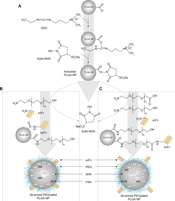
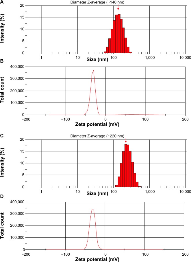
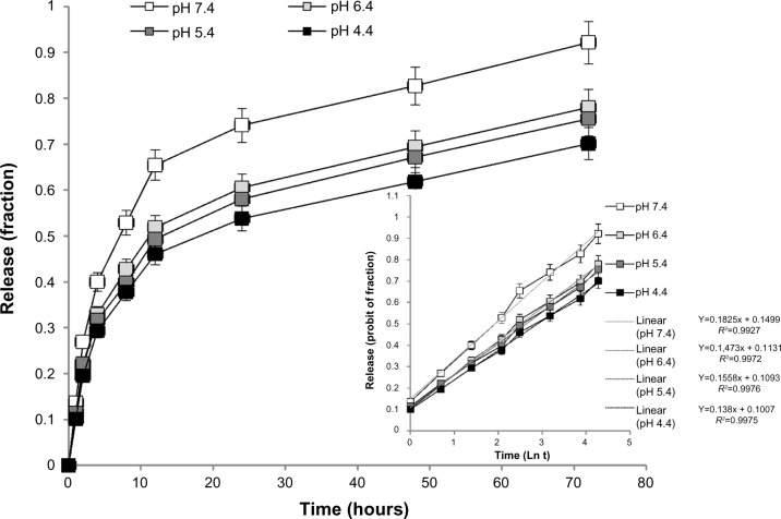
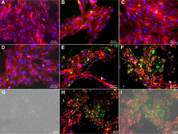
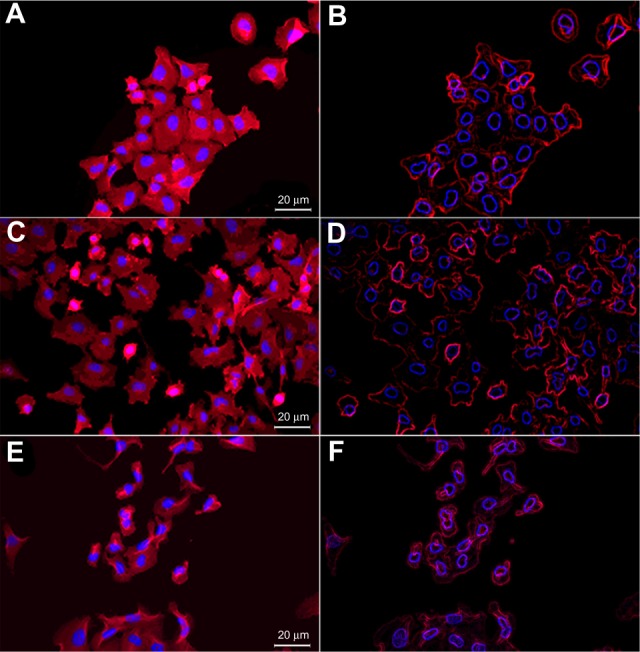
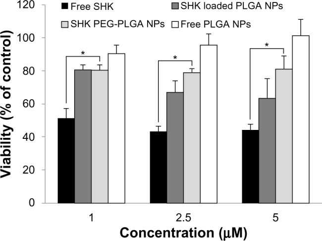
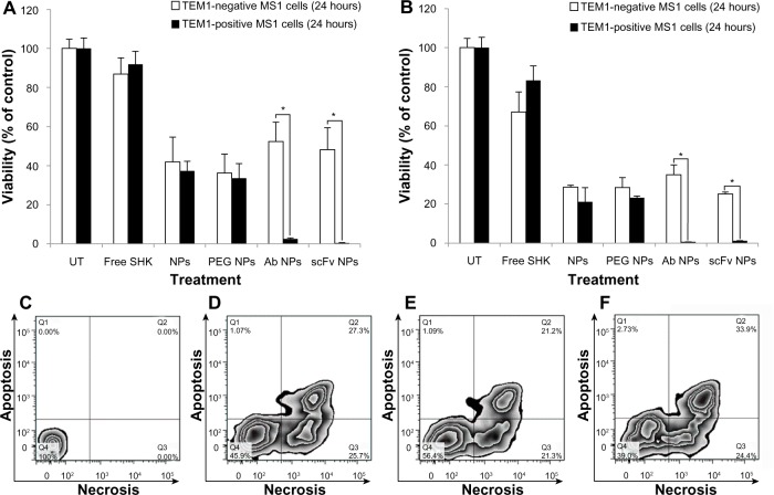
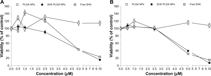
Conventional chemotherapy of ovarian cancer often fails because of initiation of drug resistance and/or side effects and trace of untouched remaining cancerous cells. This highlights an urgent need for advanced targeted therapies for effective remediation of the disease using a cytotoxic agent with immunomodulatory effects, such as shikonin (SHK). Based on preliminary experiments, we found SHK to be profoundly toxic in ovarian epithelial cancer cells (OVCAR-5 and ID8 cells) as well as in normal ovarian IOSE-398 cells, endothelial MS1 cells, and lymphocytes. To limit its cytotoxic impact solely to tumor cells within the tumor microenvironment (TME), we aimed to engineer SHK as polymeric nanoparticles (NPs) with targeting moiety toward tumor microvasculature. To this end, using single/double emulsion solvent evaporation/diffusion technique with sonication, we formulated biodegradable NPs of poly(lactic-co-glycolic acid) (PLGA) loaded with SHK. The surface of NPs was further decorated with solubilizing agent polyethylene glycol (PEG) and tumor endothelial marker 1 (TEM1)/endosialin-targeting antibody (Ab) through carbodiimide/N-hydroxysuccinimide chemistry. Having characterized the physicochemical and morphological properties of NPs, we studied their drug-release profiles using various kinetic models. The biological impact of NPs was also evaluated in tumor-associated endothelial MS1 cells, primary lymphocytes, and epithelial ovarian cancer OVCAR-5 cells. Based on particle size analysis and electron microscopy, the engineered NPs showed a smooth spherical shape with size range of 120 to 250 nm and zeta potential value of -30 to -40 mV. Drug entrapment efficiency was ~80%-90%, which was reduced to ~50%-60% upon surface decoration with PEG and Ab. The liberation of SHK from NPs showed a sustained-release profile that was best fitted with Wagner log-probability model. Fluorescence microscopy and flow cytometry analysis showed active interaction of Ab-armed NPs with TEM1-positive MS1 cells, but not with TEM1-negative MS1 cells. While exposure of the PEGylated NPs for 2 hours was not toxic to lymphocytes, long-term exposure of the Ab-armed and PEGylated NPs was significantly toxic to TEM1-positive MS1 cells and OVCAR-5 cells. Based on these findings, we propose SHK-loaded Ab-armed PEGylated PLGA NPs as a novel nanomedicine for targeted therapy of solid tumors.
卵巢癌的传统化疗常常失败,原因在于出现耐药性和/或副作用,以及残留未被清除的癌细胞。这凸显了迫切需要先进的靶向治疗方法,以使用具有免疫调节作用的细胞毒性药物(如紫草素,SHK)有效治疗该疾病。基于初步实验,我们发现SHK对卵巢上皮癌细胞(OVCAR - 5和ID8细胞)以及正常卵巢IOSE - 398细胞、内皮MS1细胞和淋巴细胞具有显著毒性。为了将其细胞毒性作用仅局限于肿瘤微环境(TME)中的肿瘤细胞,我们旨在将SHK设计成具有靶向肿瘤微脉管系统部分的聚合物纳米颗粒(NPs)。为此,我们采用单/双乳液溶剂蒸发/扩散技术并结合超声处理,制备了负载SHK的聚乳酸 - 乙醇酸共聚物(PLGA)可生物降解纳米颗粒。通过碳二亚胺/N - 羟基琥珀酰亚胺化学方法,纳米颗粒的表面进一步用增溶剂聚乙二醇(PEG)和肿瘤内皮标志物1(TEM1)/内涎蛋白靶向抗体(Ab)进行修饰。在对纳米颗粒的物理化学和形态学性质进行表征后,我们使用各种动力学模型研究了它们的药物释放曲线。还在肿瘤相关内皮MS1细胞、原代淋巴细胞和上皮性卵巢癌OVCAR - 5细胞中评估了纳米颗粒的生物学影响。基于粒径分析和电子显微镜观察,工程化纳米颗粒呈现光滑的球形,尺寸范围为120至250 nm,zeta电位值为 - 30至 - 40 mV。药物包封率约为80% - 90%,在用PEG和Ab进行表面修饰后降至约50% - 60%。SHK从纳米颗粒中的释放呈现持续释放曲线,最符合Wagner对数概率模型。荧光显微镜和流式细胞术分析表明,携带抗体的纳米颗粒与TEM1阳性MS1细胞有活跃相互作用,但与TEM1阴性MS1细胞无相互作用。虽然聚乙二醇化纳米颗粒暴露2小时对淋巴细胞无毒,但携带抗体和聚乙二醇化的纳米颗粒长期暴露对TEM1阳性MS1细胞和OVCAR - 5细胞具有显著毒性。基于这些发现,我们提出负载SHK的携带抗体聚乙二醇化PLGA纳米颗粒作为一种用于实体瘤靶向治疗的新型纳米药物。