European Commission-Joint Research Centre, Institute for Health and Consumer Protection, Ispra (VA), Italy.
European Commission-Joint Research Centre, Institute for Environment and Sustainability, Ispra (VA), Italy; FU-Berlin, Fachbereich Biologie, Chemie, Pharmazie, Berlin, Germany.
PLoS One. 2014 May 5;9(5):e96078. doi: 10.1371/journal.pone.0096078. eCollection 2014.
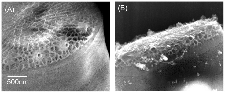
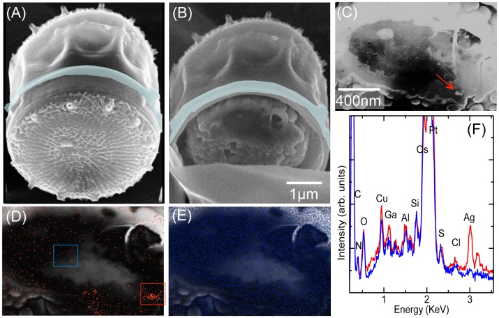
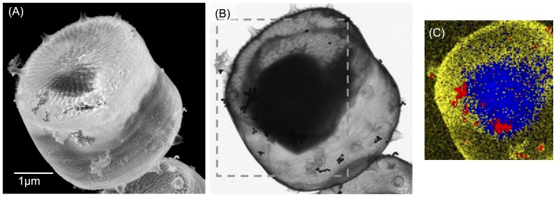
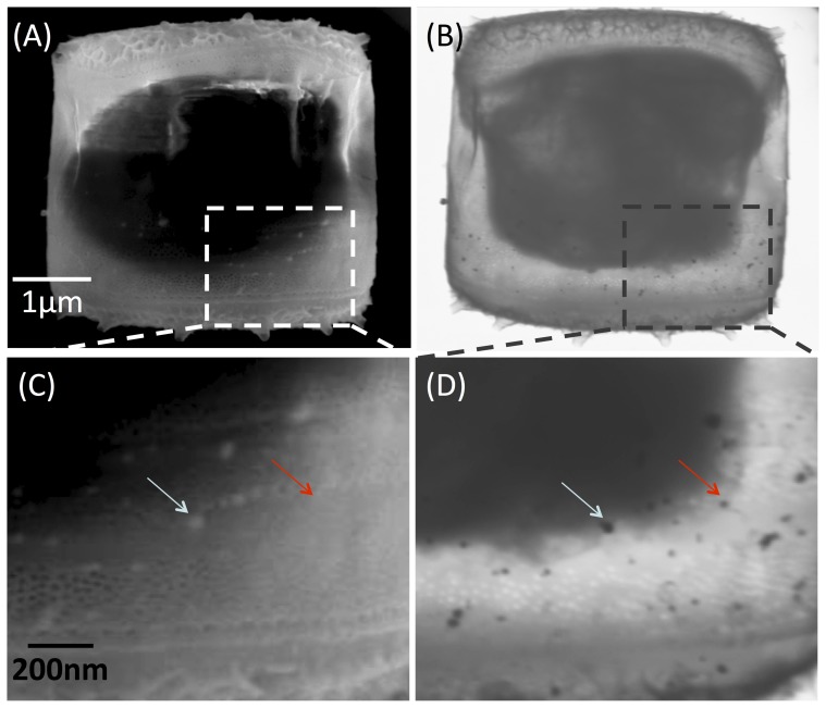
In the following article an electron/ion microscopy study will be presented which investigates the uptake of silver nanoparticles (AgNPs) by the marine diatom Thalassiosira pseudonana, a primary producer aquatic species. This organism has a characteristic silica exoskeleton that may represent a barrier for the uptake of some chemical pollutants, including nanoparticles (NPs), but that presents a technical challenge when attempting to use electron-microscopy (EM) methods to study NP uptake. Here we present a convenient method to detect the NPs interacting with the diatom cell. It is based on a fixation procedure involving critical point drying which, without prior slicing of the cell, allows its inspection using transmission electron microscopy. Employing a combination of electron and ion microscopy techniques to selectively cut the cell where the NPs were detected, we are able to demonstrate and visualize for the first time the presence of AgNPs inside the cell membrane.
以下文章将介绍一项电子/离子显微镜研究,该研究调查了海洋硅藻拟菱形藻对银纳米颗粒(AgNPs)的摄取情况,拟菱形藻是一种主要的水生初级生产者。这种生物有一个特征性的硅外壳,这可能是一些化学污染物(包括纳米颗粒(NPs))摄取的障碍,但当试图使用电子显微镜(EM)方法研究 NP 摄取时,这也带来了技术挑战。在这里,我们提出了一种方便的方法来检测与硅藻细胞相互作用的 NPs。它基于一种涉及临界点干燥的固定程序,该程序无需预先对细胞进行切片,即可使用透射电子显微镜进行检查。我们采用电子和离子显微镜技术的组合,选择性地切割检测到 NPs 的细胞,从而首次能够证明和可视化 AgNPs 存在于细胞膜内。