Hsieh Ya-Ju, Hwu Luen, Ke Chien-Chih, Yeh Skye Hsin-Hsien, Lin Chien-Feng, Chen Fu-Du, Wang Hsin-Ell, Lin Kang-Ping, Chen Ran-Chou, Liu Ren-Shyan
Department of Medical Imaging and Radiological Sciences, Kaohsiung Medical University, No. 100, Shih-Chuan 1st Road, Kaohsiung 80708, Taiwan.
Molecular and Genetic Imaging Core/Taiwan Mouse Clinic, National Comprehensive Mouse Phenotyping and Drug Testing Center, No. 201, Section 2, Shih-Pai Road, Taipei 11217, Taiwan.
Biomed Res Int. 2014;2014:605358. doi: 10.1155/2014/605358. Epub 2014 Apr 7.
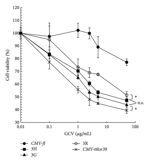
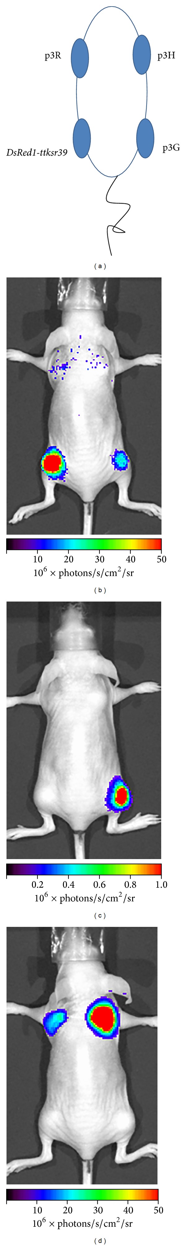
Multimodality imaging using noncytotoxic triple fusion (TF) reporter genes is an important application for cell-based tracking, drug screening, and therapy. The firefly luciferase (fl), monomeric red fluorescence protein (mrfp), and truncated herpes simplex virus type 1 thymidine kinase SR39 mutant (ttksr39) were fused together to create TF reporter gene constructs with different order. The enzymatic activities of TF protein in vitro and in vivo were determined by luciferase reporter assay, H-FEAU cellular uptake experiment, bioluminescence imaging, and micropositron emission tomography (microPET). The TF construct expressed in H1299 cells possesses luciferase activity and red fluorescence. The tTKSR39 activity is preserved in TF protein and mediates high levels of H-FEAU accumulation and significant cell death from ganciclovir (GCV) prodrug activation. In living animals, the luciferase and tTKSR39 activities of TF protein have also been successfully validated by multimodality imaging systems. The red fluorescence signal is relatively weak for in vivo imaging but may expedite FACS-based selection of TF reporter expressing cells. We have developed an optimized triple fusion reporter construct DsRedm-fl-ttksr39 for more effective and sensitive in vivo animal imaging using fluorescence, bioluminescence, and PET imaging modalities, which may facilitate different fields of biomedical research and applications.
使用无细胞毒性的三融合(TF)报告基因的多模态成像在基于细胞的追踪、药物筛选和治疗中是一项重要应用。将萤火虫荧光素酶(fl)、单体红色荧光蛋白(mrfp)和截短的单纯疱疹病毒1型胸苷激酶SR39突变体(ttksr39)融合在一起,构建了不同顺序的TF报告基因构建体。通过荧光素酶报告基因检测、H-FEAU细胞摄取实验、生物发光成像和微型正电子发射断层扫描(microPET)测定TF蛋白在体外和体内的酶活性。在H1299细胞中表达的TF构建体具有荧光素酶活性和红色荧光。tTKSR39活性在TF蛋白中得以保留,并介导高水平的H-FEAU积累以及由更昔洛韦(GCV)前药激活导致的显著细胞死亡。在活体动物中,TF蛋白的荧光素酶和tTKSR39活性也已通过多模态成像系统成功验证。红色荧光信号在体内成像时相对较弱,但可能会加快基于荧光激活细胞分选(FACS)的TF报告基因表达细胞的筛选。我们已经开发出一种优化的三融合报告基因构建体DsRedm-fl-ttksr39,用于使用荧光、生物发光和PET成像模式进行更有效和灵敏的体内动物成像,这可能会促进生物医学研究和应用的不同领域的发展。