Department of Experimental Pneumology and Allergology, Saarland University Faculty of Medicine, Kirrberger Strasse, Geb, 61,4, Homburg, Germany.
Respir Res. 2014 Jun 30;15(1):73. doi: 10.1186/1465-9921-15-73.
A neuroimmune crosstalk between dendritic cells (DCs) and airway nerves in the lung has recently been reported. However, the presence of DCs in airway sensory ganglia under normal and allergic conditions has not been explored so far. Therefore, this study aims to investigate the localisation, distribution and proliferation of DCs in airway sensory ganglia under allergic airway inflammation.
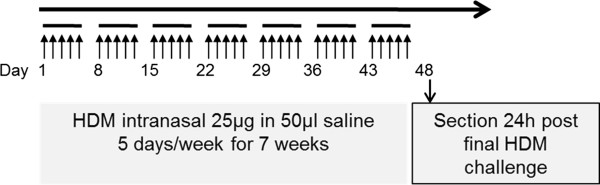
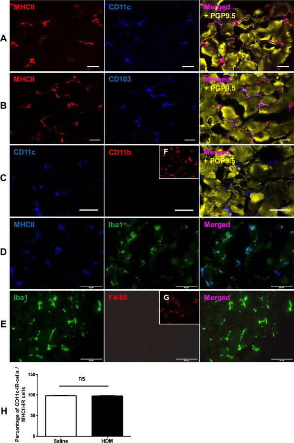
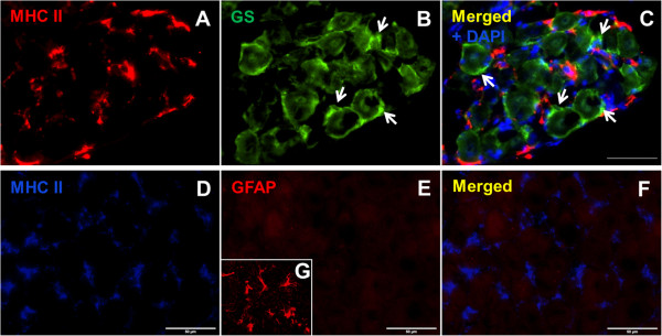
Using the house dust mite (HDM) model for allergic airway inflammation BALB/c mice were exposed to HDM extract intranasally (25 μg/50 μl) for 5 consecutive days a week over 7 weeks. With the help of the immunohistochemistry, vagal jugular-nodose ganglia complex (JNC) sections were analysed regarding their expression of DC-markers (MHC II, CD11c, CD103), the neuronal marker PGP 9.5 and the neuropeptide calcitonin gene-related peptide (CGRP) and glutamine synthetase (GS) as a marker for satellite glia cells (SGCs). To address the original source of DCs in sensory ganglia, a proliferation experiment was also carried in this study.
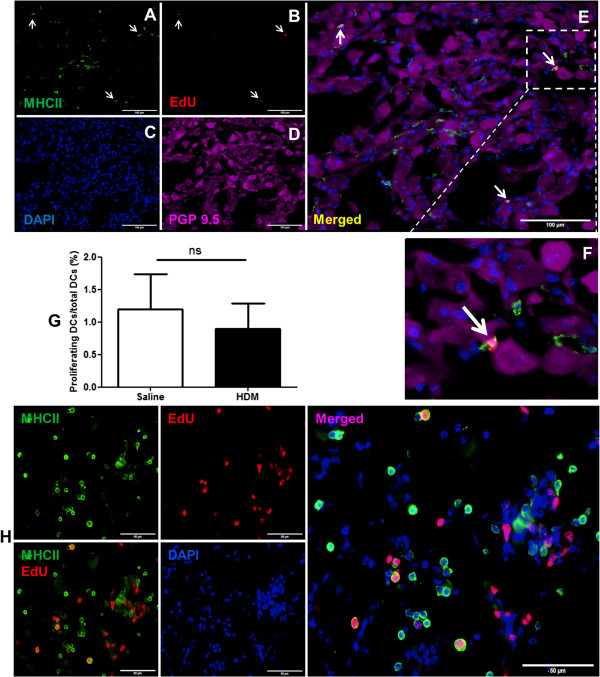
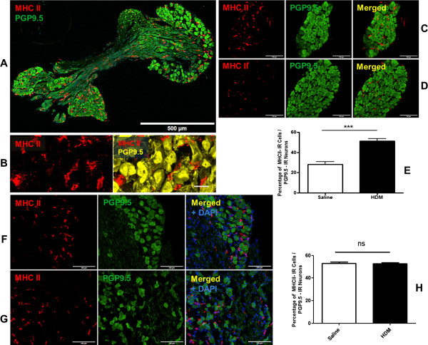
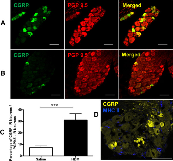
Immune cells with characteristic DC-phenotype were found to be closely located to SGCs and vagal sensory neurons under physiological conditions. The percentage of DCs in relation to neurons was significantly increased by allergic airway inflammation in comparison to the controls (HDM 51.38 ± 2.38% vs. control 28.16 ± 2.86%, p < 0.001). The present study also demonstrated that DCs were shown to proliferate in jugular-nodose ganglia, however, the proliferation rate of DCs is not significantly changed in the two treated animal groups (proliferating DCs/ total DCs: HDM 0.89 ± 0.38%, vs. control 1.19 ± 0.54%, p = 0.68). Also, increased number of CGRP-positive neurons was found in JNC after allergic sensitisation and challenge (HDM 31.16 ± 5.41% vs. control 7.16 ± 1.53%, p < 0.001).
The present findings suggest that DCs may migrate from outside into the ganglia to interact with sensory neurons enhancing or protecting the allergic airway inflammation. The increase of DCs as well as CGRP-positive neurons in airway ganglia by allergic airway inflammation indicate that intraganglionic DCs and neurons expressing CGRP may contribute to the pathogenesis of bronchial asthma. To understand this neuroimmune interaction in allergic airway inflammation further functional experiments should be carried out in future studies.
树突状细胞(DCs)与肺部气道神经之间的神经免疫相互作用最近已被报道。然而,在正常和过敏条件下,气道感觉神经节中是否存在 DCs 尚未得到探索。因此,本研究旨在探讨过敏性气道炎症下气道感觉神经节中 DCs 的定位、分布和增殖。
使用屋尘螨(HDM)模型,将 BALB/c 小鼠用 HDM 提取物经鼻腔(25μg/50μl)连续 5 天/周,每周 5 天,共 7 周。通过免疫组织化学分析,分析迷走神经颈结神经节复合体(JNC)中 DC 标志物(MHC II、CD11c、CD103)、神经元标志物 PGP 9.5、神经肽降钙素基因相关肽(CGRP)和谷氨酰胺合成酶(GS)的表达,作为卫星胶质细胞(SGCs)的标志物。为了研究感觉神经节中 DC 的起源,本研究还进行了增殖实验。
在生理条件下,发现具有典型 DC 表型的免疫细胞与 SGCs 和迷走感觉神经元紧密相邻。与对照组相比,过敏性气道炎症时 DC 占神经元的比例显著增加(HDM 51.38±2.38% vs. 对照组 28.16±2.86%,p<0.001)。本研究还表明,JNC 中的 DC 能够增殖,但在两个处理组的 DC 增殖率没有明显变化(增殖的 DC/总 DC:HDM 0.89±0.38%,vs. 对照组 1.19±0.54%,p=0.68)。此外,过敏致敏和激发后 JNC 中 CGRP 阳性神经元的数量也增加(HDM 31.16±5.41% vs. 对照组 7.16±1.53%,p<0.001)。
本研究结果表明,DC 可能从外部迁移到神经节中与感觉神经元相互作用,从而增强或保护过敏性气道炎症。过敏性气道炎症引起的气道神经节中 DCs 和 CGRP 阳性神经元的增加表明,神经节内的 DCs 和表达 CGRP 的神经元可能参与了支气管哮喘的发病机制。为了进一步了解过敏性气道炎症中的神经免疫相互作用,未来的研究应进行更多的功能实验。