Heinrich Pette Institute, Leibniz Institute for Experimental Virology , Hamburg , Germany.
Institute for Tumor Biology, University Hospital Eppendorf , Hamburg , Germany.
Front Oncol. 2014 Jun 26;4:168. doi: 10.3389/fonc.2014.00168. eCollection 2014.
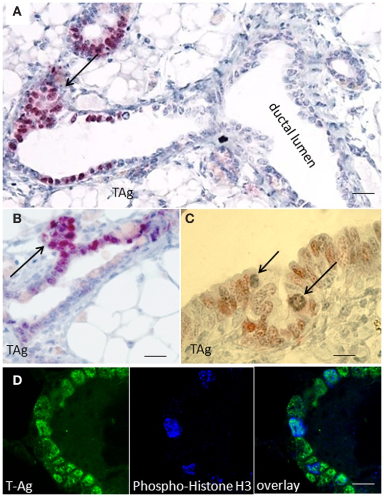
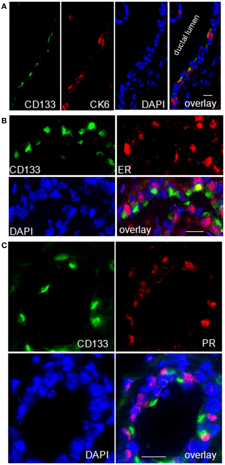
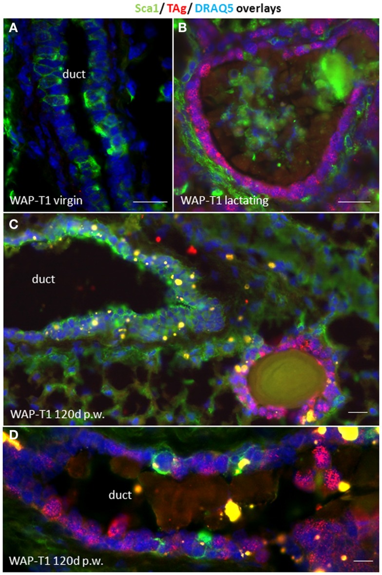
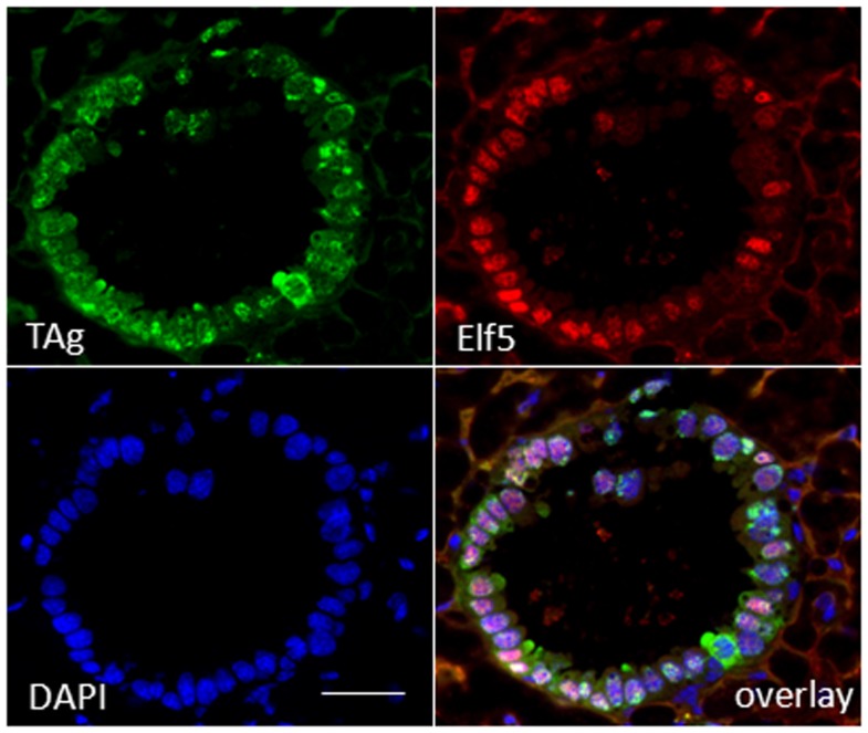
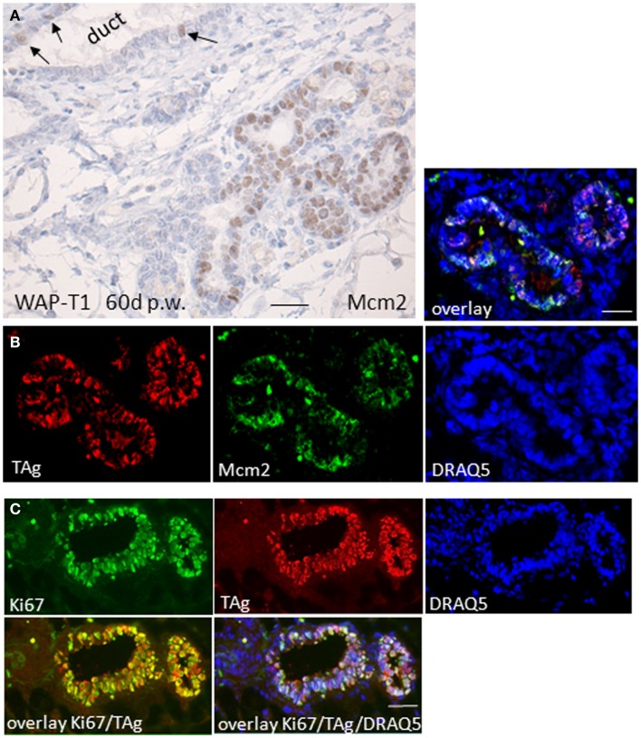
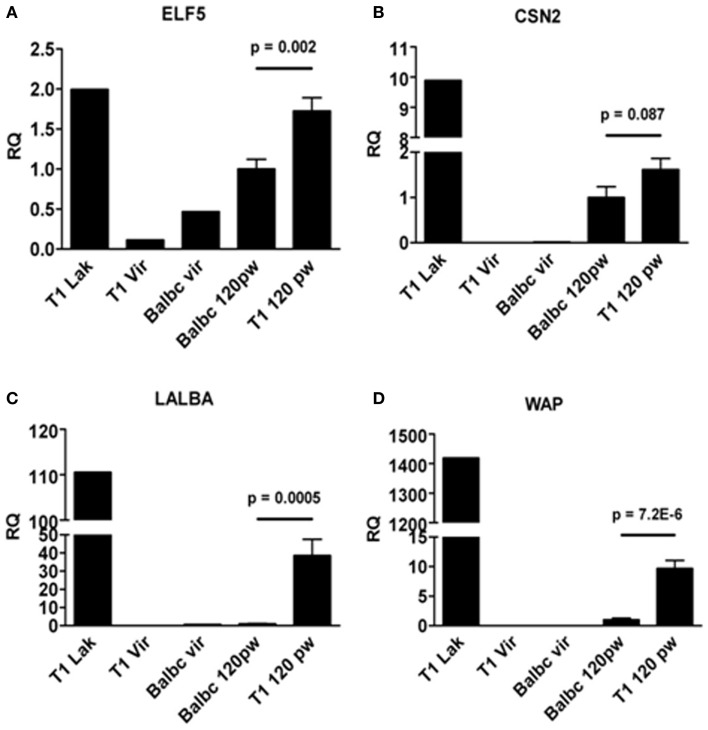
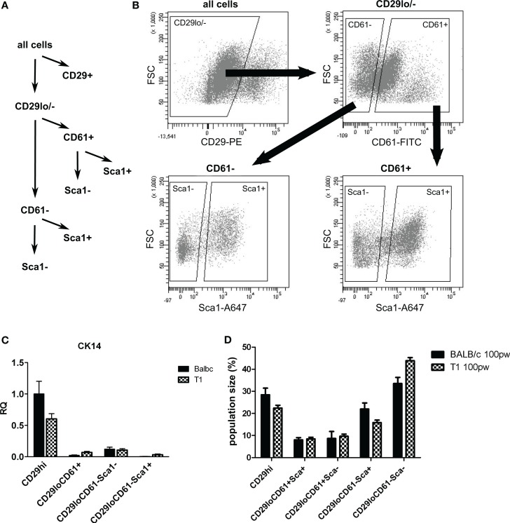
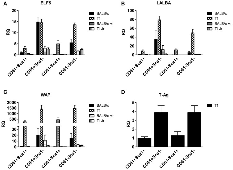
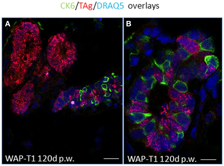
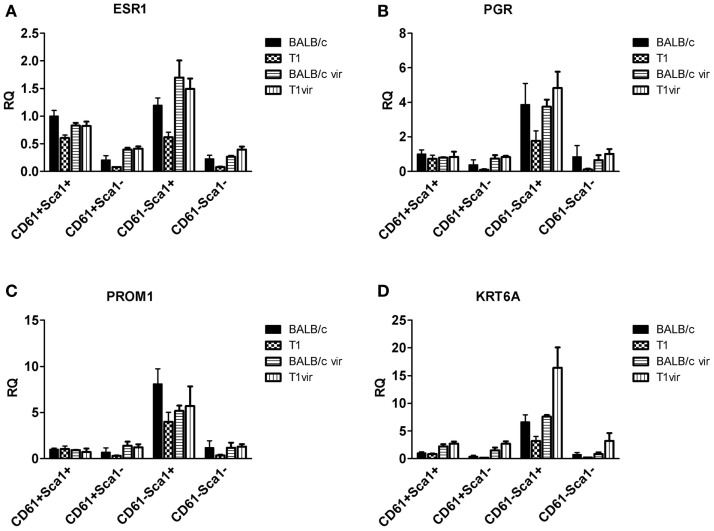
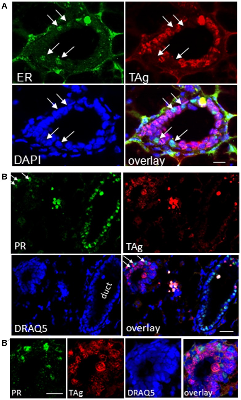
WAP-T1 transgenic mice express SV40-TAg under control of the whey acidic protein (WAP) promoter, which directs activity of this strong viral oncogene to luminal cells of the mammary gland. Resting uniparous WAP-T1 glands develop hyperplasia composed of TAg positive cells prior to appearance of advanced tumor stages. We show that cells in hyperplasia display markers of alveolar differentiation, suggesting that TAg targets differentiating cells of the alveolar compartment. The glands show significant expression of Elf5 and milk genes (Lalba, Csn2, and Wap). TAg expressing cells largely co-stain with antibodies to Elf5, lack the epithelial marker Sca1, and are hormone receptor negative. High expression levels of Elf5 but not of milk genes are also seen in resting glands of normal BALB/c mice. This indicates that expression of Elf5 in resting WAP-T1 glands is not specifically induced by TAg. CK6a positive luminal cells lack TAg. These cells co-express the markers prominin-1, CK6a, and Sca1, and are positive for hormone receptors. These hormone sensitive cells localize to ducts and seem not to be targeted by TAg. Despite reaching an advanced stage in alveolar differentiation, the cells in hyperplasia do not exit the cell cycle. Thus, expression of TAg in conjunction with regular morphogenetic processes of alveologenesis seem to provide the basis for a hormone independent, unscheduled proliferation of differentiating cells in resting glands of WAP-T1 transgenic mice, leading to the formation of hyperplastic lesions.
WAP-T1 转基因小鼠在乳清酸性蛋白 (WAP) 启动子的控制下表达 SV40-TAg,该启动子将这种强病毒癌基因的活性定向到乳腺的腔细胞。静止的单胎 WAP-T1 腺体会在出现晚期肿瘤阶段之前,由 TAg 阳性细胞组成增生。我们表明,增生中的细胞显示出肺泡分化的标志物,表明 TAg 靶向肺泡隔的分化细胞。这些腺体显示出 Elf5 和乳基因(Lalba、Csn2 和 Wap)的显著表达。表达 TAg 的细胞与 Elf5 抗体的染色大部分重叠,缺乏上皮标志物 Sca1,并且激素受体阴性。在正常 BALB/c 小鼠的静止 WAP-T1 腺体内也观察到 Elf5 的高表达水平,但缺乏乳基因。这表明静止 WAP-T1 腺体内 Elf5 的表达不是由 TAg 特异性诱导的。CK6a 阳性腔细胞不含有 TAg。这些细胞共同表达标志物 prominin-1、CK6a 和 Sca1,并对激素受体呈阳性。这些激素敏感细胞定位于导管,似乎不受 TAg 的靶向作用。尽管在肺泡分化中达到晚期阶段,增生中的细胞仍未退出细胞周期。因此,TAg 的表达与肺泡发生的常规形态发生过程相结合,似乎为静止的 WAP-T1 转基因小鼠乳腺中分化细胞的激素独立、不定期增殖提供了基础,导致增生性病变的形成。