Plesner Annette, Ten Holder Joris T, Verchere C Bruce
Departments of Pathology and Laboratory Medicine, Child & Family Research Institute, University of British Columbia, Vancouver, British Columbia, Canada.
Departments of Pathology and Laboratory Medicine, Child & Family Research Institute, University of British Columbia, Vancouver, British Columbia, Canada; Department of Surgery, Child & Family Research Institute, University of British Columbia, Vancouver, British Columbia, Canada.
PLoS One. 2014 Aug 7;9(8):e102843. doi: 10.1371/journal.pone.0102843. eCollection 2014.
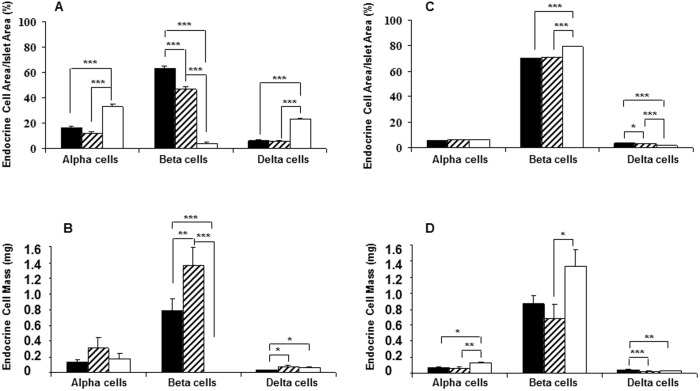
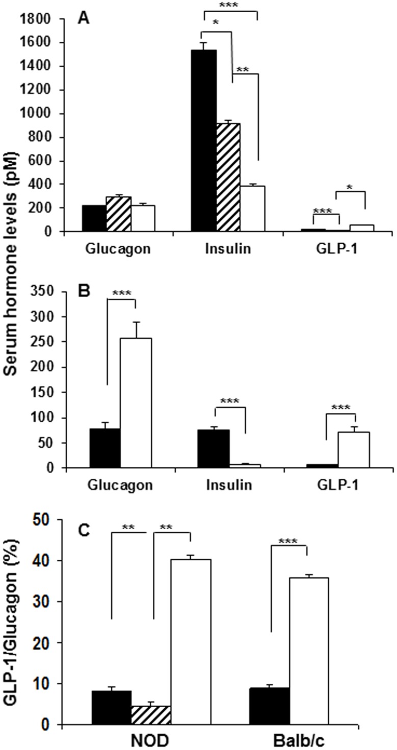
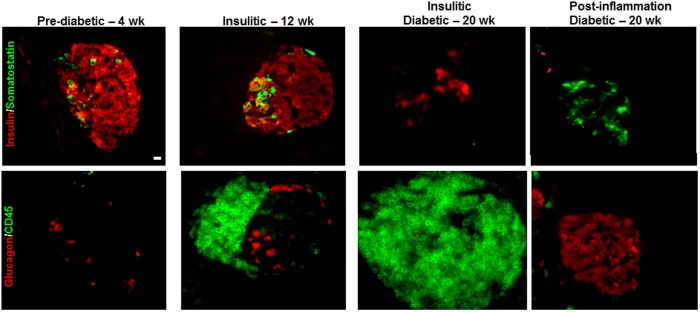
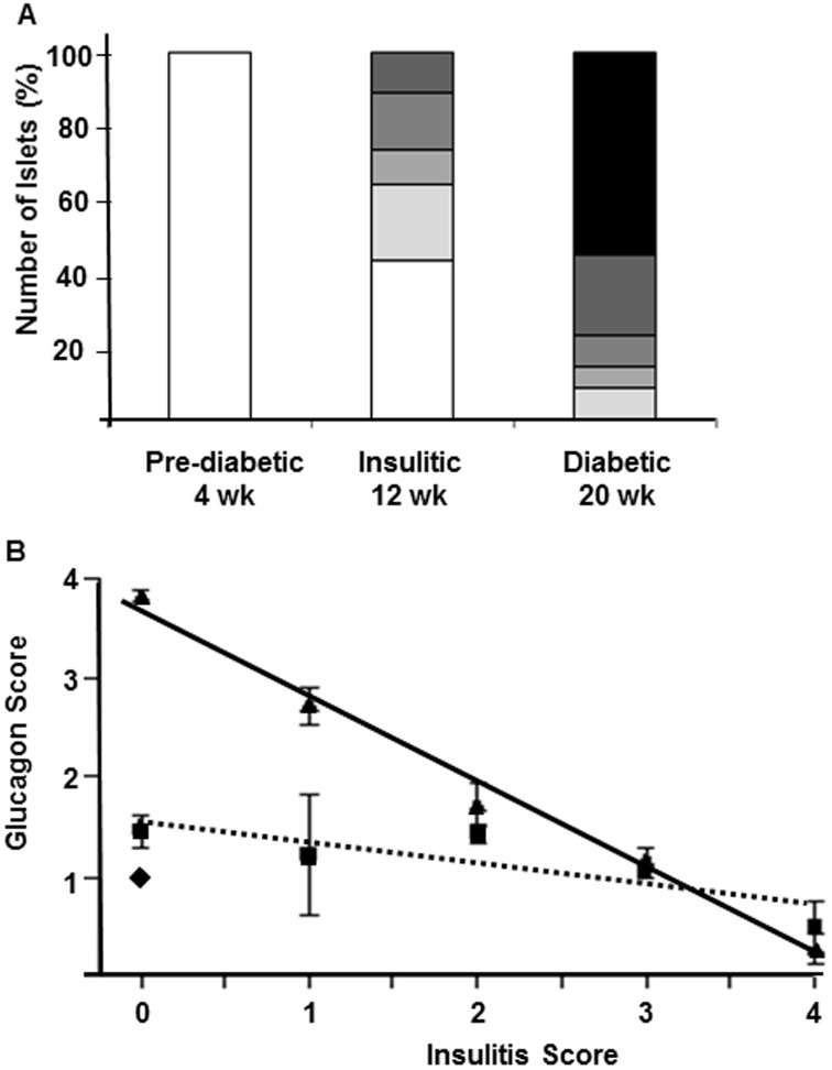
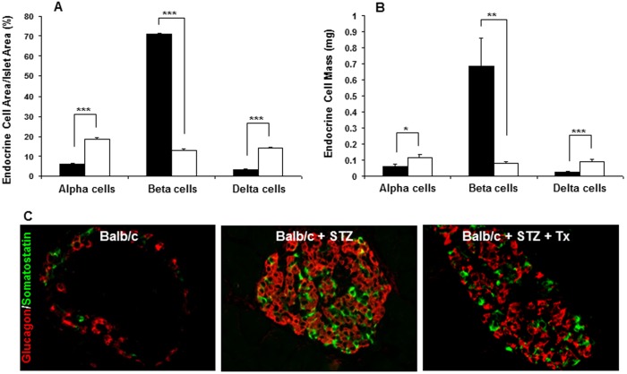
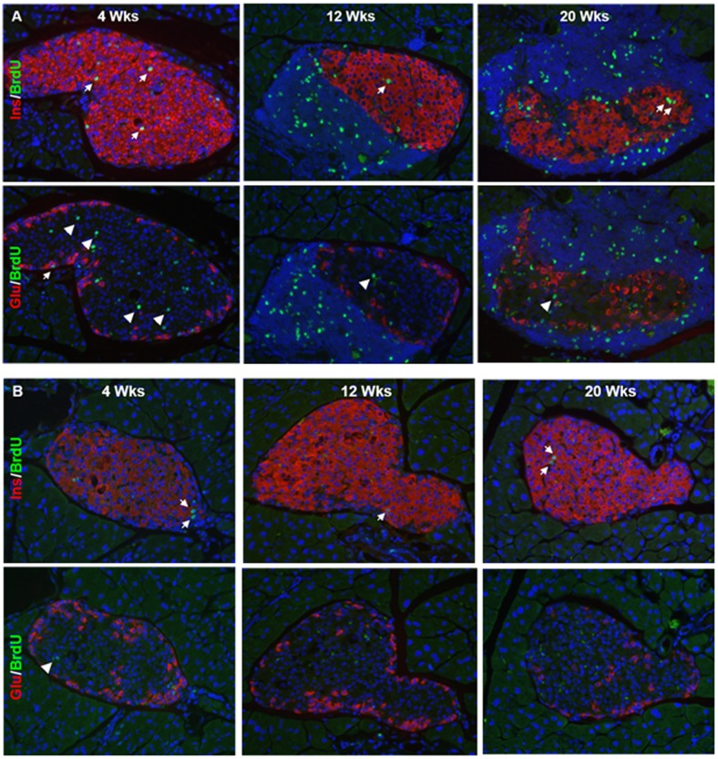
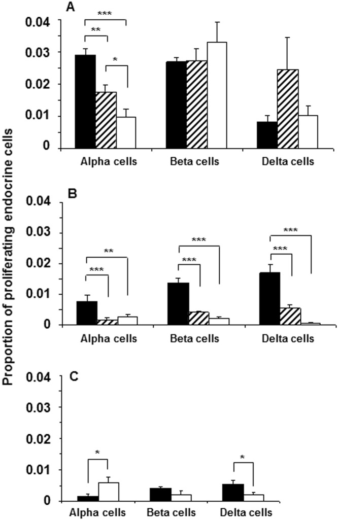
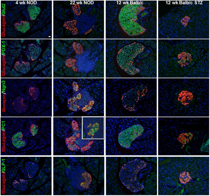
Islet alpha- and delta-cells are spared autoimmune destruction directed at beta-cells in type 1 diabetes resulting in an apparent increase of non-beta endocrine cells in the islet core. We determined how islet remodeling in autoimmune diabetes compares to streptozotocin (STZ)-induced diabetes. Islet cell mass, proliferation, and immune cell infiltration in pancreas sections from diabetic NOD mice and mice with STZ-induced diabetes was assessed using quantitative image analysis. Serial sections were stained for various beta-cell markers and Ngn3, typically restricted to embryonic tissue, was only upregulated in diabetic NOD mouse islets. Serum levels of insulin, glucagon and GLP-1 were measured to compare hormone levels with respect to disease state. Total pancreatic alpha-cell mass did not change as autoimmune diabetes developed in NOD mice despite the proportion of islet area comprised of alpha- and delta-cells increased. By contrast, alpha- and delta-cell mass was increased in mice with STZ-induced diabetes. Serum levels of glucagon reflected these changes in alpha-cell mass: glucagon levels remained constant in NOD mice over time but increased significantly in STZ-induced diabetes. Increased serum GLP-1 levels were found in both models of diabetes, likely due to alpha-cell expression of prohormone convertase 1/3. Alpha- or delta-cell mass in STZ-diabetic mice did not normalize by replacement of insulin via osmotic mini-pumps or islet transplantation. Hence, the inflammatory milieu in NOD mouse islets may restrict alpha-cell expansion highlighting important differences between these two diabetes models and raising the possibility that increased alpha-cell mass might contribute to the hyperglycemia observed in the STZ model.
在1型糖尿病中,胰岛α细胞和δ细胞免受针对β细胞的自身免疫破坏,导致胰岛核心中非β内分泌细胞明显增加。我们确定了自身免疫性糖尿病中的胰岛重塑与链脲佐菌素(STZ)诱导的糖尿病相比如何。使用定量图像分析评估糖尿病NOD小鼠和STZ诱导的糖尿病小鼠胰腺切片中的胰岛细胞质量、增殖和免疫细胞浸润。对连续切片进行各种β细胞标志物染色,通常仅限于胚胎组织的Ngn3仅在糖尿病NOD小鼠胰岛中上调。测量血清胰岛素、胰高血糖素和GLP-1水平以比较疾病状态下的激素水平。尽管由α细胞和δ细胞组成的胰岛面积比例增加,但随着NOD小鼠自身免疫性糖尿病的发展,胰腺α细胞总质量并未改变。相比之下,STZ诱导的糖尿病小鼠的α细胞和δ细胞质量增加。胰高血糖素血清水平反映了α细胞质量的这些变化:随着时间的推移,NOD小鼠的胰高血糖素水平保持恒定,但在STZ诱导的糖尿病中显著增加。在两种糖尿病模型中均发现血清GLP-1水平升高,这可能是由于激素原转化酶1/3在α细胞中的表达。通过渗透微型泵或胰岛移植补充胰岛素后,STZ糖尿病小鼠的α细胞或δ细胞质量并未恢复正常。因此,NOD小鼠胰岛中的炎症环境可能会限制α细胞的扩张,突出了这两种糖尿病模型之间的重要差异,并增加了α细胞质量增加可能导致STZ模型中观察到的高血糖的可能性。