Lee Tsung-Ming, Lin Shinn-Zong, Chang Nen-Chung
Department of Medicine, Cardiology Section, China Medical University-An Nan Hospital, Tainan, Taiwan; Department of Medicine, China Medical University, Taichung, Taiwan; Department of Internal Medicine, School of Medicine, College of Medicine, Taipei Medical University, Taipei, Taiwan.
J Cell Mol Med. 2014 Dec;18(12):2454-65. doi: 10.1111/jcmm.12430. Epub 2014 Sep 25.
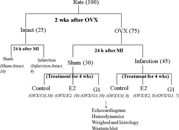
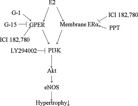
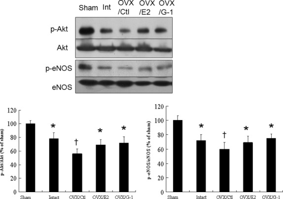
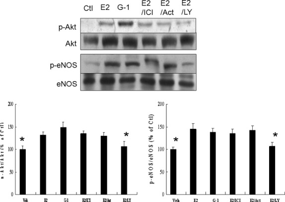
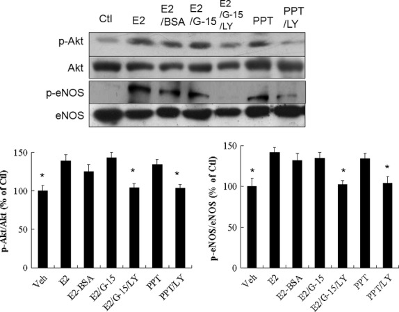
Clinical and experimental studies have established that gender is a factor in the development of ventricular hypertrophy. We investigated whether the attenuated hypertrophic effect of oestradiol was via activation of phosphatidylinositol 3-kinase (PI3K)/Akt/endothelial nitric oxide synthase (eNOS) through non-genomic action. Twenty-four hours after coronary ligation, female Wistar rats were randomized into control, subcutaneous oestradiol treatment or a G-protein coupled oestrogen receptor (GPER) agonist, G-1 and treated for 4 weeks starting from 2 weeks after bilateral ovariectomy. Ventricular hypertrophy assessed by cardiomyocyte size after infarction was similarly attenuated by oestradiol or G-1 in infarcted rats. The phosphorylation of Akt and eNOS was significantly decreased in infarcted rats and restored by oestradiol and G-1, implying the GPER pathway in this process. Oestradiol-induced Akt phosphorylation was not abrogated by G-15 (a GPER blocker). Akt activation was not inhibited by actinomycin D. When a membrane-impermeable oestrogen-albumin construct was applied, similar responses in terms of eNOS activation to those of oestradiol were achieved. Furthermore, PPT, an ERα receptor agonist, activated the phosphorylation of Akt and eNOS. Thus, membrane ERα receptor played a role in mediating the phosphorylation of Akt and eNOS. The specific PI3K inhibitor, LY290042, completely abolished Akt activation and eNOS phosphorylation in infarcted hearts treated with either oestradiol or oestradiol + G-15. These data support the conclusions that oestradiol improves ventricular remodelling by both GPER- and membrane-bound ERα-dependent mechanisms that converge into the PI3K/Akt/eNOS pathway, unveiling a novel mechanism by which oestradiol regulates pathological cardiomyocyte growth after infarction.
临床和实验研究已证实性别是心室肥大发展的一个因素。我们研究了雌二醇的肥大效应减弱是否是通过非基因组作用激活磷脂酰肌醇3激酶(PI3K)/蛋白激酶B(Akt)/内皮型一氧化氮合酶(eNOS)来实现的。冠状动脉结扎24小时后,将雌性Wistar大鼠随机分为对照组、皮下注射雌二醇治疗组或G蛋白偶联雌激素受体(GPER)激动剂G-1组,并在双侧卵巢切除术后2周开始治疗4周。梗死大鼠中,通过梗死心肌细胞大小评估的心室肥大,经雌二醇或G-1治疗后同样减轻。梗死大鼠中Akt和eNOS的磷酸化显著降低,经雌二醇和G-1治疗后恢复,这意味着在此过程中存在GPER途径。G-15(一种GPER阻滞剂)并未消除雌二醇诱导的Akt磷酸化。放线菌素D未抑制Akt激活。当应用一种不能透过细胞膜的雌激素-白蛋白构建体时,在eNOS激活方面获得了与雌二醇相似的反应。此外,雌激素受体α(ERα)激动剂PPT激活了Akt和eNOS的磷酸化。因此,膜ERα受体在介导Akt和eNOS的磷酸化中发挥了作用。特异性PI3K抑制剂LY290042完全消除了用雌二醇或雌二醇+G-15治疗的梗死心脏中Akt激活和eNOS磷酸化。这些数据支持以下结论:雌二醇通过GPER和膜结合ERα依赖性机制改善心室重构,这两种机制汇聚到PI3K/Akt/eNOS途径,揭示了雌二醇调节梗死后病理性心肌细胞生长的新机制。