Raghavendra Srinidhi Surya, Hindlekar Ajit Narayan, Desai Niranjan Nanasaheb, Vyavahare Nishant Kishor, Napte Bandu Devrao
Department of Conservative Dentistry and Endodontics, Sinhgad Dental College and Hospital, Pune, Maharashtra, India.
Indian J Dent. 2014 Jul;5(3):152-6. doi: 10.4103/0975-962X.140837.
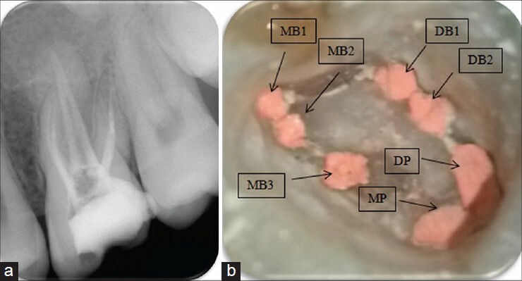
The main objective of root canal treatment is thorough cleaning and shaping of the entire pulp space and its complete filling with an inert filling material. A major cause of post-treatment disease is the inability to locate, debride or adequately fill all canals of the root canal system. The form, configuration, and number of root canals in the maxillary first molars have been discussed for more than half a century. Maxillary first molars commonly present with three roots and three canals, with a second mesiobuccal canal (MB2) also present. With the advent of improved magnification there are reports of multiple root canals in the maxillary first molars. Nonsurgical endodontic therapy of a left maxillary first molar with three roots and seven root canals was successfully performed under a dental operating microscope. The diagnosis of multiple root canals was confirmed with the help of Cone Beam Computed Tomography (CBCT) images.
根管治疗的主要目标是对整个牙髓腔进行彻底清洁和塑形,并用惰性填充材料将其完全充填。治疗后疾病的一个主要原因是无法定位、清创或充分充填根管系统的所有根管。上颌第一磨牙根管的形态、结构和数量已被讨论了半个多世纪。上颌第一磨牙通常有三个牙根和三个根管,也存在第二近中颊根管(MB2)。随着放大技术的改进,有报道称上颌第一磨牙存在多个根管。在牙科手术显微镜下成功地对上颌左侧第一磨牙(有三个牙根和七个根管)进行了非手术牙髓治疗。借助锥形束计算机断层扫描(CBCT)图像证实了多根管的诊断。