Trojsi Francesca, Caiazzo Giuseppina, Corbo Daniele, Piccirillo Giovanni, Cristillo Viviana, Femiano Cinzia, Ferrantino Teresa, Cirillo Mario, Monsurrò Maria Rosaria, Esposito Fabrizio, Tedeschi Gioacchino
Department of Medical, Surgical, Neurological, Metabolic and Aging Sciences, Second University of Naples, Naples, Italy; MRI Research Center SUN-FISM-Second University of Naples, 80138 Naples, Italy.
Department of Neuroscience, University of Parma, 43100 Parma, Italy.
PLoS One. 2015 Mar 20;10(3):e0119045. doi: 10.1371/journal.pone.0119045. eCollection 2015.
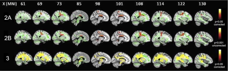
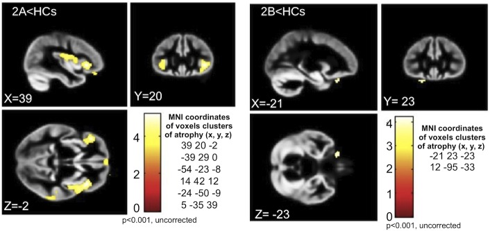
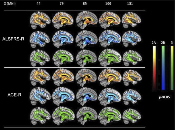
Neurodegenerative process in amyotrophic lateral sclerosis (ALS) has been proven to involve several cortical and subcortical brain regions within and beyond motor areas. However, how ALS pathology spreads progressively during disease evolution is still unknown. In this cross-sectional study we investigated 54 ALS patients, divided into 3 subsets according to the clinical stage, and 18 age and sex-matched healthy controls, by using tract-based spatial statistics (TBSS) diffusion tensor imaging (DTI) and voxel-based morphometry (VBM) analyses. We aimed to identify white (WM) and gray matter (GM) patterns of disease distinctive of each clinical stage, corresponding to specific clinical milestones. ALS cases in stage 2A (i.e., at diagnosis) were characterized by GM and WM impairment of left motor and premotor cortices and brainstem at ponto-mesenchephalic junction. ALS patients in clinical stage 2B (with impairment of two functional regions) exhibited decreased fractional anisotropy (FA) (p<0.001, uncorrected) and increased mean (MD) and radial diffusivity (RD) (p<0.001, uncorrected) in the left cerebellar hemisphere and brainstem precerebellar nuclei, as well as in motor areas, while GM atrophy (p<0.001, uncorrected) was detected only in the left inferior frontal gyrus and right cuneus. Finally, ALS patients in stage 3 (with impairment of three functional regions) exhibited decreased FA and increased MD and RD (p<0.05, corrected) within WM underneath bilateral pre and postcentral gyri, corpus callosum midbody, long associative tracts and midbrain, while no significant clusters of GM atrophy were observed. Our findings reinforce the hypothesis that the neurodegenerative process propagates along the axonal pathways and develops beyond motor areas from early stages, involving progressively several frontotemporal regions and their afferents and efferents, while the detection of GM atrophy in earlier stages and its disappearance in later stages may be the result of reactive gliosis.
肌萎缩侧索硬化症(ALS)的神经退行性过程已被证明涉及运动区域内外的几个皮质和皮质下脑区。然而,ALS病理在疾病发展过程中如何逐渐扩散仍不清楚。在这项横断面研究中,我们使用基于纤维束的空间统计学(TBSS)扩散张量成像(DTI)和基于体素的形态计量学(VBM)分析,对54例ALS患者(根据临床阶段分为3个亚组)和18名年龄和性别匹配的健康对照进行了研究。我们旨在确定每个临床阶段特有的疾病白质(WM)和灰质(GM)模式,这些模式对应于特定的临床里程碑。2A期(即诊断时)的ALS病例表现为左侧运动和运动前皮质以及脑桥中脑交界处脑干的GM和WM损伤。临床2B期(有两个功能区域受损)的ALS患者在左侧小脑半球、脑桥小脑前核以及运动区域表现出分数各向异性(FA)降低(p<0.001,未校正),平均扩散率(MD)和径向扩散率(RD)增加(p<0.001,未校正),而GM萎缩仅在左侧额下回和右侧楔叶被检测到(p<0.001,未校正)。最后,3期(有三个功能区域受损)的ALS患者在双侧中央前回和中央后回下方的WM、胼胝体中部、长联合纤维束和中脑内表现出FA降低,MD和RD增加(p<0.05,校正),而未观察到明显的GM萎缩簇。我们的研究结果强化了这样一种假设,即神经退行性过程沿着轴突通路传播,从早期就超出运动区域发展,逐渐累及几个额颞区域及其传入和传出纤维,而早期GM萎缩的检测及其在后期的消失可能是反应性胶质增生的结果。