Zhu Dan, Xie Li, Karimian Negar, Liang Tao, Kang Youhou, Huang Ya-Chi, Gaisano Herbert Y
Department of Medicine, Faculty of Medicine, University of Toronto, Toronto, ON, Canada.
Mol Metab. 2015 Feb 16;4(5):418-26. doi: 10.1016/j.molmet.2015.02.004. eCollection 2015 May.
Pancreatic beta-cells express three Munc18 isoforms. Much is known about the roles of Munc18a (pre-docked secretory granules-SGs) and Munc18b (newcomer SGs and SG-SG fusion) in insulin exocytosis. Although shown to influence glucose-stimulated insulin secretion (GSIS) in rodents the precise role of Munc18c in insulin SG exocytosis has not been elucidated. We here examined the role of Munc18c in human pancreatic beta-cells.
Munc18c-shRNA/RFP lenti-virus (versus control virus) was used to knock down the expression level of Munc18c in human islets or single beta-cells. Insulin secretion and granule exocytosis were measured by performing islets perifusion, single-cell patch clamp capacitance measurements and total internal reflection fluorescence microscopy (TIRFM).
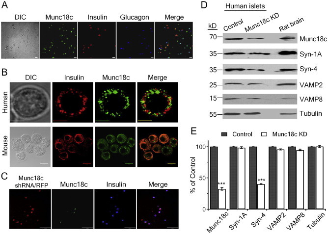
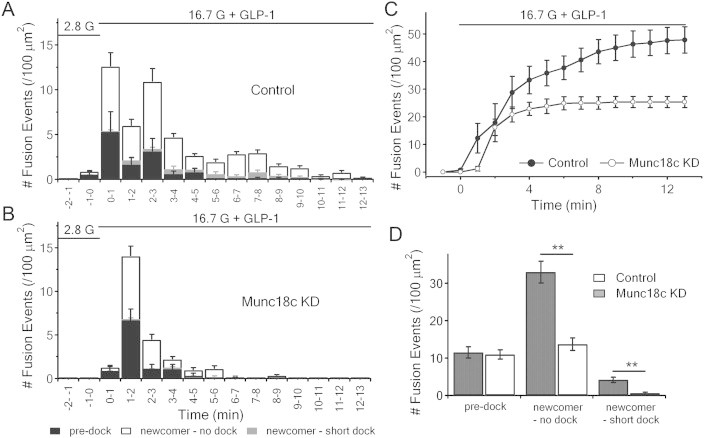
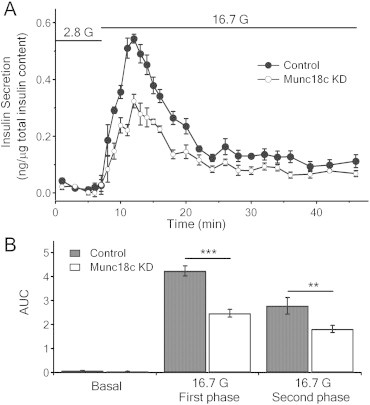
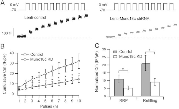
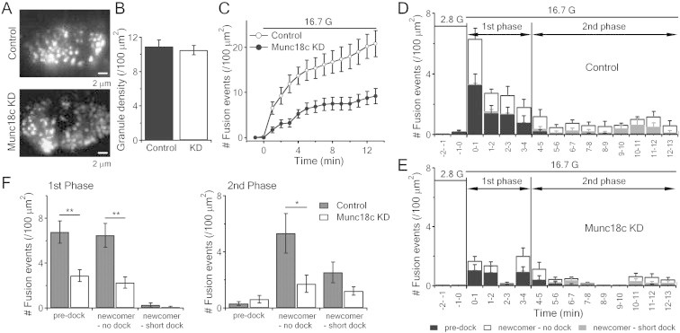
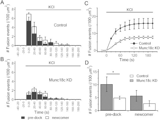
Munc18c is most abundant in the cytosol of human beta-cells. Endogenous function of Munc18c was assessed by knocking down (KD) its islet expression by 70% employing lenti-shRNA virus. Munc18c-KD caused reduction in cognate syntaxin-4 islet expression but not in other exocytotic proteins, resulting in the reduction in GSIS in first- (by 42%) and second phases (by 35%). Single cell analyses of RFP-tagged Munc18c-KD beta-cells by patch clamp capacitance measurements showed inhibition in both readily-releasable pool (by 52%) and mobilization from the reserve pool (by 57%). TIRFM to assess single SG behavior showed that Munc18c-KD inhibition of first phase GSIS was attributed to reduction in exocytosis of pre-docked and newcomer SGs, and second phase inhibition attributed entirely to reduction in newcomer SG fusion (SGs that undergo minimal residence or docking time at the plasma membrane before fusion).
Munc18c is involved in the distinct molecular machineries that affect exocytosis of both predocked and newcomer SG pools that underlie Munc18c's role in first and second phases of GSIS, respectively.
胰腺β细胞表达三种Munc18亚型。关于Munc18a(预对接分泌颗粒-SGs)和Munc18b(新形成的SGs和SG-SG融合)在胰岛素胞吐作用中的作用,我们已了解很多。尽管已证明Munc18c在啮齿动物中会影响葡萄糖刺激的胰岛素分泌(GSIS),但其在胰岛素SG胞吐作用中的精确作用尚未阐明。我们在此研究了Munc18c在人胰腺β细胞中的作用。
使用Munc18c-shRNA/RFP慢病毒(与对照病毒相比)来敲低人胰岛或单个β细胞中Munc18c的表达水平。通过进行胰岛灌流、单细胞膜片钳电容测量和全内反射荧光显微镜(TIRFM)来测量胰岛素分泌和颗粒胞吐作用。
Munc18c在人β细胞的胞质溶胶中含量最为丰富。通过使用慢病毒-shRNA病毒将其在胰岛中的表达敲低70%来评估Munc18c的内源性功能。Munc18c敲低导致同源Syntaxin-4在胰岛中的表达降低,但其他胞吐蛋白的表达未受影响,从而导致第一阶段(降低42%)和第二阶段(降低35%)的GSIS降低。通过膜片钳电容测量对带有RFP标签的Munc18c敲低β细胞进行单细胞分析显示,可快速释放池(降低52%)和储备池的动员(降低57%)均受到抑制。TIRFM用于评估单个SG行为,结果显示Munc18c敲低对第一阶段GSIS的抑制作用归因于预对接和新形成的SGs胞吐作用的降低,而第二阶段的抑制作用完全归因于新形成的SG融合(融合前在质膜停留或对接时间最短的SGs)的降低。
Munc18c参与了不同的分子机制,这些机制分别影响预对接和新形成的SG池的胞吐作用,这也是Munc18c在GSIS第一阶段和第二阶段发挥作用的基础。