Matz Joachim M, Goosmann Christian, Brinkmann Volker, Grützke Josephine, Ingmundson Alyssa, Matuschewski Kai, Kooij Taco W A
1] Parasitology Unit, Max Planck Institute for Infection Biology, Charitéplatz 1, 10117 Berlin, Germany [2] Institute of Biology, Humboldt University, 10117 Berlin, Germany [3] Department of Medical Microbiology, Radboud Institute for Molecular Life Sciences, Radboud University Medical Centre, P.O. Box 9101, 6500 HB Nijmegen, The Netherlands.
Microscopy Core Facility, Max Planck Institute for Infection Biology, Charitéplatz 1, 10117 Berlin, Germany.
Sci Rep. 2015 Jul 29;5:12532. doi: 10.1038/srep12532.
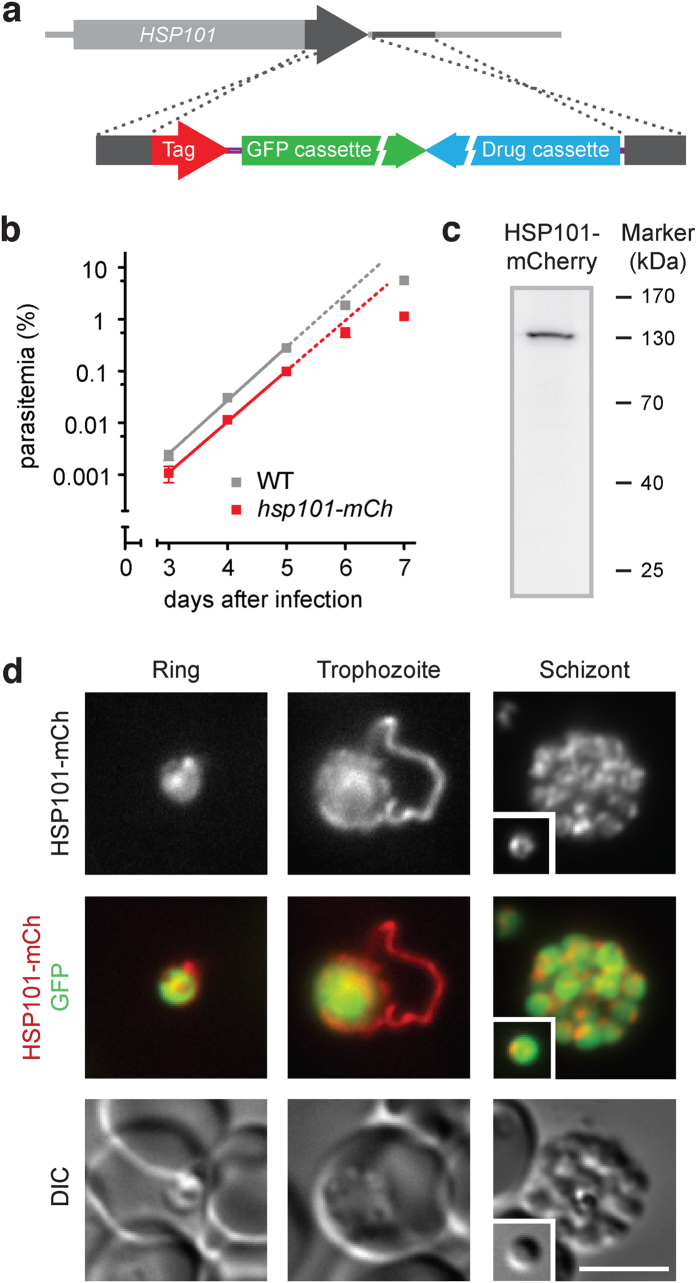
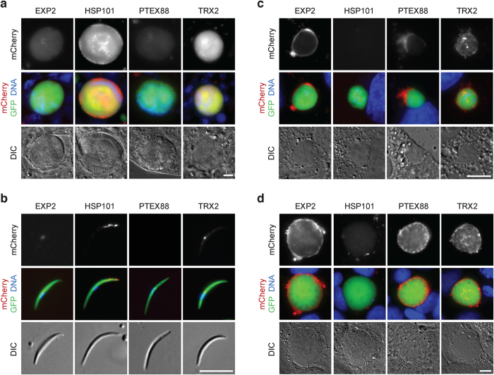
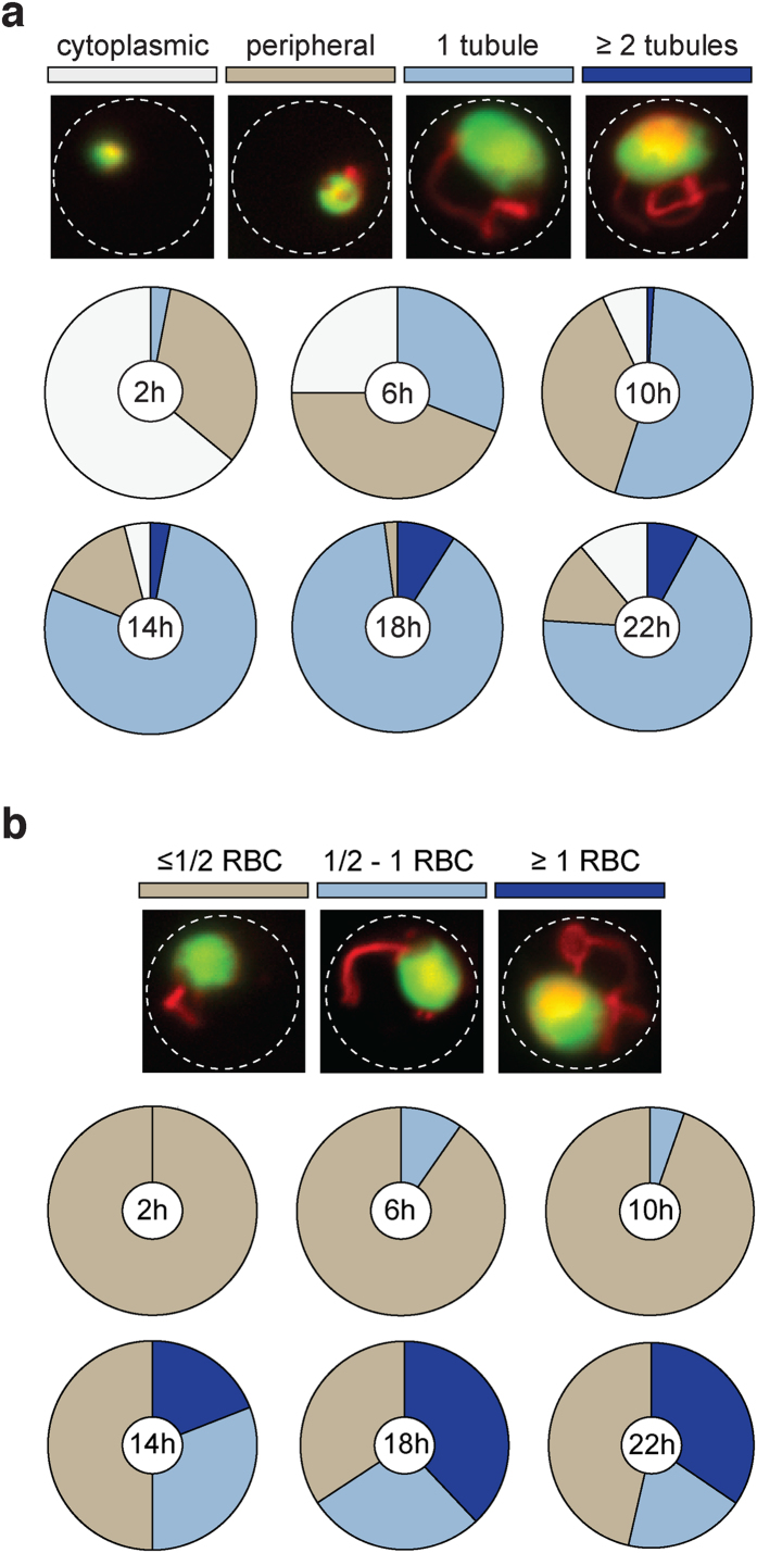
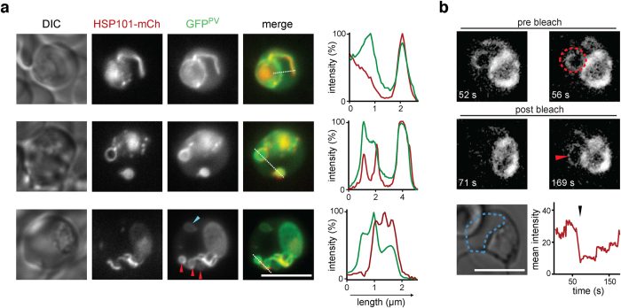
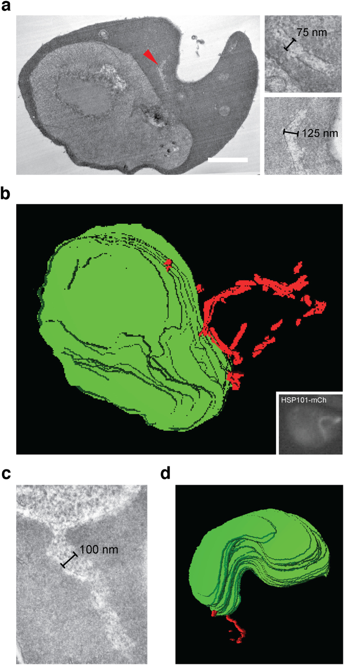
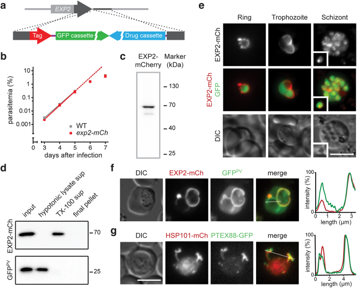
The erythrocyte is an extraordinary host cell for intracellular pathogens and requires extensive remodelling to become permissive for infection. Malaria parasites modify their host red blood cells through protein export to acquire nutrients and evade immune responses. Endogenous fluorescent tagging of three signature proteins of the Plasmodium berghei translocon of exported proteins (PTEX), heat shock protein 101, exported protein 2 (EXP2), and PTEX88, revealed motile, tubular extensions of the parasitophorous vacuole that protrude from the parasite far into the red blood cell. EXP2 displays a more prominent presence at the periphery of the parasite, consistent with its proposed role in pore formation. The tubular compartment is most prominent during trophozoite growth. Distinct spatiotemporal expression of individual PTEX components during sporogony and liver-stage development indicates additional functions and tight regulation of the PTEX translocon during parasite life cycle progression. Together, live cell imaging and correlative light and electron microscopy permitted previously unrecognized spatiotemporal and subcellular resolution of PTEX-containing tubules in murine malaria parasites. These findings further refine current models for Plasmodium-induced erythrocyte makeover.
红细胞是细胞内病原体的特殊宿主细胞,需要进行广泛重塑才能允许感染。疟原虫通过蛋白质输出修饰其宿主红细胞,以获取营养并逃避免疫反应。对伯氏疟原虫输出蛋白转运体(PTEX)的三种标志性蛋白、热休克蛋白101、输出蛋白2(EXP2)和PTEX88进行内源性荧光标记,发现寄生泡有能动的管状延伸,从寄生虫延伸到红细胞深处。EXP2在寄生虫周边的存在更为显著,与其在孔形成中的假定作用一致。管状区室在滋养体生长期间最为突出。在孢子生殖和肝期发育过程中,各个PTEX成分的独特时空表达表明在寄生虫生命周期进展过程中,PTEX转运体具有额外功能且受到严格调控。活细胞成像以及相关的光学和电子显微镜技术相结合,使得在鼠疟原虫中对含PTEX的小管进行了前所未有的时空和亚细胞分辨率观察。这些发现进一步完善了当前疟原虫诱导红细胞重塑的模型。