Kuramoto Kunitaka, Beppu Toru, Namimoto Tomohiro, Hayashi Hiromitsu, Imai Katsunori, Nitta Hidetoshi, Hashimoto Daisuke, Chikamoto Akira, Ishiko Takatoshi, Iyama Ken-Ichi, Ikeda Osamu, Yamashita Yasuyuki, Baba Hideo
Department of Gastroenterological Surgery, Graduate School of Life Sciences, Kumamoto University, 1-1-1 Honjo, Kumamoto City, Kumamoto 860-8556 Japan.
Department of Gastroenterological Surgery, Graduate School of Life Sciences, Kumamoto University, 1-1-1 Honjo, Kumamoto City, Kumamoto 860-8556 Japan ; Department of Multidisciplinary Treatment for Gastroenterological Cancer, Kumamoto University Hospital, 1-1-1 Honjo, Kumamoto City, Kumamoto 860-8556 Japan.
Surg Case Rep. 2015;1(1):38. doi: 10.1186/s40792-015-0038-0. Epub 2015 Apr 24.
Angiomyolipoma is a unique mesenchymal neoplasm composed of blood vessels as well as smooth muscle and adipose cells. The liver is a less common site of origin, and hepatic angiomyolipoma is often an incidental finding on diagnostic imaging or is identified on evaluation of nonspecific symptoms.
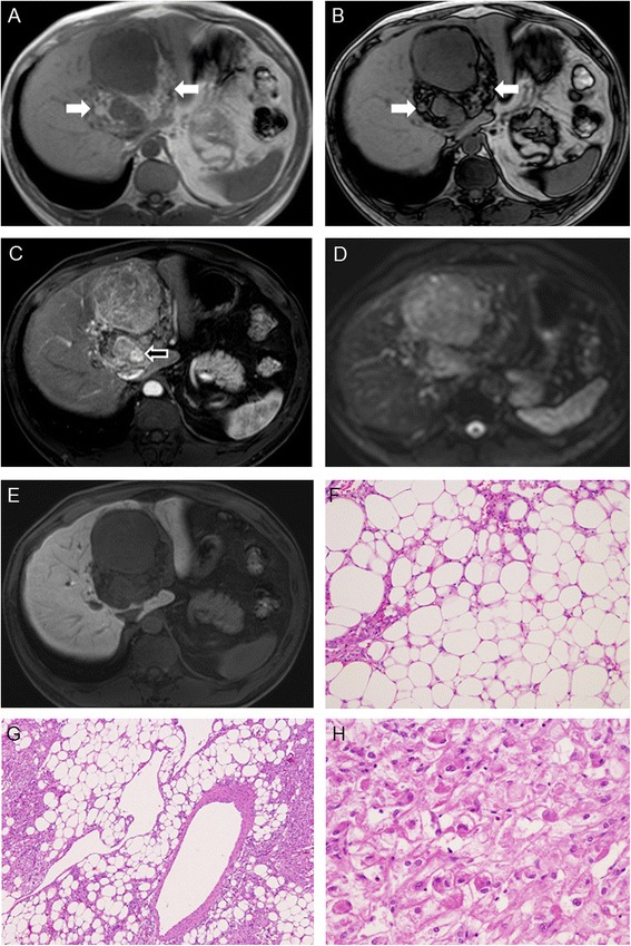
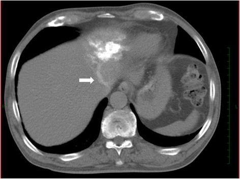
We experienced four patients who were diagnosed histologically with hepatic angiomyolipoma. The preoperative diagnoses were angiomyolipoma in two patients, hepatocellular carcinoma in one, and cavernous hemangioma in one. Three patients were treated with hepatectomy (one laparoscopic and two open approaches), and the diagnosis was completed by histological investigation of the resected specimen. The remaining one was diagnosed from tumor needle biopsy. Diffusion-weighted magnetic resonance imaging (MRI) with respiratory triggering using b values of 0 and 800 s/mm(2) was employed. An apparent diffusion coefficient map was generated from b values of 0 and 800 s/mm(2) for calculation of the apparent diffusion coefficient. The apparent diffusion coefficient values were calculated as 3.66, 1.21, 1.80, and 0.91 in patients 1 to 4, respectively. In MRI imaging, fat component was clearly demonstrated with chemical shift imaging in three patients. Early venous return was detected in three patients with computed tomography angiography.
Fat component and early venous return are important for a correct diagnosis of hepatic angiomyolipoma. Unfortunately, apparent diffusion coefficient values in hepatic angiomyolipoma were overlapping with those in other benign and malignant tumors.
血管平滑肌脂肪瘤是一种独特的间叶性肿瘤,由血管、平滑肌和脂肪细胞组成。肝脏是其较少见的起源部位,肝血管平滑肌脂肪瘤常在诊断性影像学检查时偶然发现,或在评估非特异性症状时被识别。
我们遇到了4例经组织学诊断为肝血管平滑肌脂肪瘤的患者。术前诊断中,2例为血管平滑肌脂肪瘤,1例为肝细胞癌,1例为海绵状血管瘤。3例患者接受了肝切除术(1例腹腔镜手术和2例开放手术),通过对切除标本的组织学检查完成诊断。其余1例通过肿瘤穿刺活检确诊。采用了b值为0和800 s/mm²的呼吸触发扩散加权磁共振成像(MRI)。根据b值为0和800 s/mm²生成表观扩散系数图以计算表观扩散系数。患者1至4的表观扩散系数值分别计算为3.66、1.21、1.80和0.91。在MRI成像中,3例患者通过化学位移成像清晰显示了脂肪成分。3例患者通过计算机断层血管造影检测到早期静脉回流。
脂肪成分和早期静脉回流对肝血管平滑肌脂肪瘤的正确诊断很重要。遗憾的是,肝血管平滑肌脂肪瘤的表观扩散系数值与其他良性和恶性肿瘤的表观扩散系数值重叠。