Jochelson Maxine S, Lebron Lizza, Jacobs Stefanie S, Zheng Junting, Moskowitz Chaya S, Powell Simon N, Sacchini Virgilio, Ulaner Gary A, Morris Elizabeth A, Dershaw D David
1 All authors: Memorial Sloan Kettering Cancer Center, 1275 York Ave, New York, NY 10065.
AJR Am J Roentgenol. 2015 Oct;205(4):899-904. doi: 10.2214/AJR.14.13804.
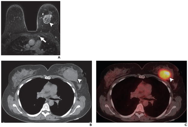
The purpose of this study was to assess the prevalence of internal mammary node (IMN) adenopathy in patients with breast cancer and compare breast MRI and PET/CT for detection of IMN adenopathy.
This retrospective study included 90 women who underwent MRI and PET/CT before neoadjuvant chemotherapy for clinical stage IIA through IIIA disease. MRI and PET/CT examinations were read independently by two readers trained in breast imaging and nuclear medicine. All patients underwent follow-up MRI at the end of chemotherapy, and 10 with hypermetabolic IMNs underwent follow-up PET/CT. Histology was not obtained. Women were considered to have IMN adenopathy when nodes seen on MRI or having standardized uptake value (SUV) greater than mediastinal blood pool decreased in either size or SUV (or both) after treatment. Features including lymphovascular invasion, tumor quadrant(s), and axillary adenopathy were compared between presence and absence of IMN adenopathy using Fisher's exact test. Prevalence was determined on the basis of the percentage of patients with IMN adenopathy by either modality. The McNemar test compared the prevalence of IMN adenopathy on MRI to its prevalence on PET/CT.
Prevalence of IMN adenopathy was 16% (14/90) by MRI and 14% (13/90) by PET/CT (p = 0.317). After chemotherapy, IMN adenopathy resolved in 12 of 14 patients (86%). In two patients with poor responses in primary tumors, IMN adenopathy persisted, and both patients developed metastatic disease within 6 months. At 3 years, survival was significantly worse in patients with IMN adenopathy than in those without (85.7% vs 53.3%, respectively; p = 0.009).
In women with advanced breast cancer receiving neoadjuvant chemo-therapy, prevalence of IMN adenopathy was 16%, equally detected by breast MRI and PET/CT. Identification of IMN adenopathy may affect treatment and provides prognostic information.
本研究旨在评估乳腺癌患者内乳淋巴结(IMN)肿大的患病率,并比较乳腺MRI和PET/CT检测IMN肿大的效果。
本回顾性研究纳入了90例临床分期为IIA至IIIA期疾病且在新辅助化疗前接受MRI和PET/CT检查的女性。MRI和PET/CT检查由两名接受过乳腺影像学和核医学培训的阅片者独立进行。所有患者在化疗结束时接受随访MRI检查,10例IMN代谢增高的患者接受随访PET/CT检查。未获取组织学结果。当MRI上显示的淋巴结或标准化摄取值(SUV)大于纵隔血池的淋巴结在治疗后大小或SUV(或两者)减小,则认为女性存在IMN肿大。使用Fisher精确检验比较有和无IMN肿大患者之间的包括淋巴管侵犯、肿瘤象限和腋窝淋巴结肿大等特征。患病率根据两种检查方式中存在IMN肿大的患者百分比来确定。McNemar检验比较了MRI上IMN肿大的患病率与其在PET/CT上的患病率。
MRI检测到IMN肿大的患病率为16%(14/90),PET/CT检测到的患病率为14%(13/90)(p = 0.317)。化疗后,14例患者中有12例(86%)的IMN肿大消失。在2例原发肿瘤反应不佳的患者中,IMN肿大持续存在,且这两名患者均在6个月内发生了转移。3年时,有IMN肿大的患者生存率明显低于无IMN肿大的患者(分别为85.7%和53.3%;p = 0.009)。
在接受新辅助化疗的晚期乳腺癌女性中,IMN肿大的患病率为16%,乳腺MRI和PET/CT对其检测效果相同。IMN肿大的识别可能会影响治疗并提供预后信息。