• 文献检索
  • 文档翻译
  • 深度研究
  • 学术资讯
  • Suppr Zotero 插件Zotero 插件
  • 邀请有礼
  • 套餐&价格
  • 历史记录
应用&插件
Suppr Zotero 插件Zotero 插件浏览器插件Mac 客户端Windows 客户端微信小程序
定价
高级版会员购买积分包购买API积分包
服务
文献检索文档翻译深度研究API 文档MCP 服务
关于我们
关于 Suppr公司介绍联系我们用户协议隐私条款
关注我们

Suppr 超能文献

核心技术专利:CN118964589B侵权必究
粤ICP备2023148730 号-1Suppr @ 2026

文献检索

告别复杂PubMed语法,用中文像聊天一样搜索,搜遍4000万医学文献。AI智能推荐,让科研检索更轻松。

立即免费搜索

文件翻译

保留排版,准确专业,支持PDF/Word/PPT等文件格式,支持 12+语言互译。

免费翻译文档

深度研究

AI帮你快速写综述,25分钟生成高质量综述,智能提取关键信息,辅助科研写作。

立即免费体验

口腔内无线线圈的牙科 MRI。

Dental MRI using wireless intraoral coils.

机构信息

Medical Physics, Department of Radiology, University Medical Center Freiburg, Freiburg, Germany.

Division of Oral and Maxillofacial Surgery, University Medical Center Freiburg, Freiburg, Germany.

出版信息

Sci Rep. 2016 Mar 29;6:23301. doi: 10.1038/srep23301.

DOI:10.1038/srep23301
PMID:27021387
原文链接:https://pmc.ncbi.nlm.nih.gov/articles/PMC4810435/
Abstract

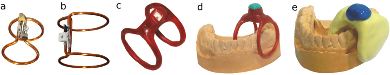
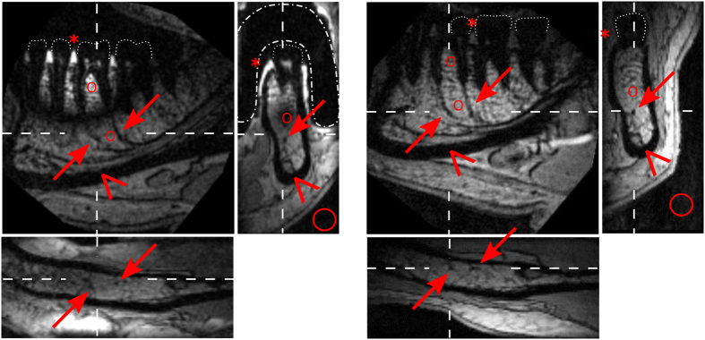
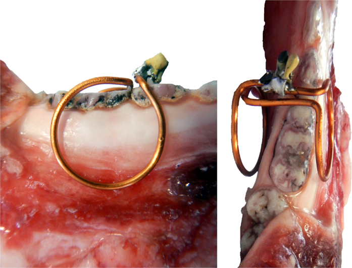
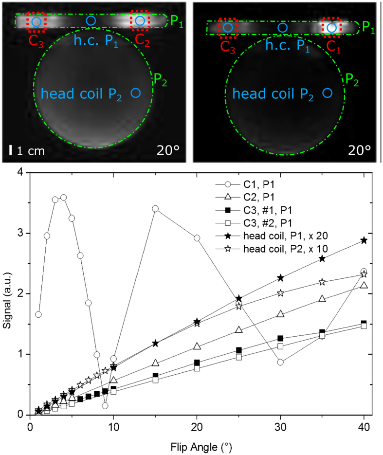
Currently, the gold standard for dental imaging is projection radiography or cone-beam computed tomography (CBCT). These methods are fast and cost-efficient, but exhibit poor soft tissue contrast and expose the patient to ionizing radiation (X-rays). The need for an alternative imaging modality e.g. for soft tissue management has stimulated a rising interest in dental magnetic resonance imaging (MRI) which provides superior soft tissue contrast. Compared to X-ray imaging, however, so far the spatial resolution of MRI is lower and the scan time is longer. In this contribution, we describe wireless, inductively-coupled intraoral coils whose local sensitivity enables high resolution MRI of dental soft tissue. In comparison to CBCT, a similar image quality with complementary contrast was obtained ex vivo. In-vivo, a voxel size of the order of 250 ∙ 250 ∙ 500 μm(3) was achieved in 4 min only. Compared to dental MRI acquired with clinical equipment, the quality of the images was superior in the sensitive volume of the coils and is expected to improve the planning of interventions and monitoring thereafter. This method may enable a more accurate dental diagnosis and avoid unnecessary interventions, improving patient welfare and bringing MRI a step closer to becoming a radiation-free alternative for dental imaging.

摘要

目前,牙科影像学的金标准是投影放射摄影或锥形束计算机断层扫描(CBCT)。这些方法快速且具有成本效益,但软组织对比度差,并使患者暴露在电离辐射(X 射线)下。需要替代的成像方式,例如软组织管理,激发了人们对牙科磁共振成像(MRI)的兴趣,它提供了更好的软组织对比度。然而,与 X 射线成像相比,到目前为止,MRI 的空间分辨率较低,扫描时间较长。在本研究中,我们描述了无线感应式口腔内线圈,其局部灵敏度能够实现高分辨率的牙科软组织 MRI。与 CBCT 相比,在离体条件下获得了具有互补对比度的相似图像质量。在体内,仅用 4 分钟即可实现约 250 × 250 × 500μm(3)的体素大小。与临床设备采集的牙科 MRI 相比,线圈敏感体积内的图像质量更好,预计将改善干预计划及其后的监测。这种方法可以实现更准确的牙科诊断,避免不必要的干预,提高患者的福利,并使 MRI 更接近成为一种无辐射的牙科影像学替代方法。

https://cdn.ncbi.nlm.nih.gov/pmc/blobs/975a/4810435/74da31a91f16/srep23301-f8.jpg
https://cdn.ncbi.nlm.nih.gov/pmc/blobs/975a/4810435/d876b1cb2be6/srep23301-f1.jpg
https://cdn.ncbi.nlm.nih.gov/pmc/blobs/975a/4810435/f8b8c43cbb32/srep23301-f2.jpg
https://cdn.ncbi.nlm.nih.gov/pmc/blobs/975a/4810435/e208350ef9b7/srep23301-f3.jpg
https://cdn.ncbi.nlm.nih.gov/pmc/blobs/975a/4810435/c52e5ce13c31/srep23301-f4.jpg
https://cdn.ncbi.nlm.nih.gov/pmc/blobs/975a/4810435/af350709dae7/srep23301-f5.jpg
https://cdn.ncbi.nlm.nih.gov/pmc/blobs/975a/4810435/868c8e01d342/srep23301-f6.jpg
https://cdn.ncbi.nlm.nih.gov/pmc/blobs/975a/4810435/9f3be9c53ed9/srep23301-f7.jpg
https://cdn.ncbi.nlm.nih.gov/pmc/blobs/975a/4810435/74da31a91f16/srep23301-f8.jpg

相似文献

1
Dental MRI using wireless intraoral coils.口腔内无线线圈的牙科 MRI。
Sci Rep. 2016 Mar 29;6:23301. doi: 10.1038/srep23301.
2
Magnetic resonance imaging of intraoral hard and soft tissues using an intraoral coil and FLASH sequences.使用口腔内线圈和快速低角度激发序列对口腔内硬组织和软组织进行磁共振成像。
Eur Radiol. 2016 Dec;26(12):4616-4623. doi: 10.1007/s00330-016-4254-1. Epub 2016 Feb 24.
3
A quality assurance framework for the fully automated and objective evaluation of image quality in cone-beam computed tomography.用于锥形束计算机断层扫描图像质量全自动客观评估的质量保证框架。
Med Phys. 2014 Mar;41(3):031901. doi: 10.1118/1.4863507.
4
Effects of Exposure Parameters and Voxel Size for Cone-Beam Computed Tomography on the Image Matching Accuracy with an Optical Dental Scan Image: An In Vitro Study.锥形束计算机断层扫描的曝光参数和体素大小对与光学牙科扫描图像的图像匹配精度的影响:一项体外研究。
Biomed Res Int. 2021 Jun 10;2021:6971828. doi: 10.1155/2021/6971828. eCollection 2021.
5
Dental magnetic resonance imaging: making the invisible visible.口腔磁共振成像:让不可见变为可见。
J Endod. 2011 Jun;37(6):745-52. doi: 10.1016/j.joen.2011.02.022. Epub 2011 Apr 6.
6
[Comparison of a dental cone beam CT with a multi-detector row CT on effective doses and physical image quality].牙科锥形束CT与多排探测器CT在有效剂量和物理图像质量方面的比较
Nihon Hoshasen Gijutsu Gakkai Zasshi. 2011;67(1):25-31. doi: 10.6009/jjrt.67.25.
7
Dental CBCT equipment and performance issues.牙科锥形束计算机断层扫描设备及性能问题。
Radiat Prot Dosimetry. 2013 Feb;153(2):212-8. doi: 10.1093/rpd/ncs289. Epub 2012 Nov 21.
8
Technical aspects of dental CBCT: state of the art.牙科锥形束计算机断层扫描的技术层面:当前技术水平
Dentomaxillofac Radiol. 2015;44(1):20140224. doi: 10.1259/dmfr.20140224.
9
The evolution and application of dental maxillofacial imaging modalities.口腔颌面成像技术的发展与应用
Dent Clin North Am. 2008 Oct;52(4):689-705, v. doi: 10.1016/j.cden.2008.05.006.
10
Mobile C-arm cone-beam CT for guidance of spine surgery: image quality, radiation dose, and integration with interventional guidance.移动式 C 臂锥形束 CT 引导脊柱手术:图像质量、辐射剂量和与介入引导的整合。
Med Phys. 2011 Aug;38(8):4563-74. doi: 10.1118/1.3597566.

引用本文的文献

1
Beyond X-Rays: Unveiling the Future of Dental Diagnosis with Dental Magnetic Resonance Imaging.超越X光:用牙科磁共振成像揭示牙科诊断的未来。
Diagnostics (Basel). 2025 May 1;15(9):1153. doi: 10.3390/diagnostics15091153.
2
Can MRI be a potential substitute for CT in cephalometric analysis for radiation-free diagnoses?在头影测量分析中,磁共振成像(MRI)能否成为计算机断层扫描(CT)的潜在替代品,用于无辐射诊断?
J Orofac Orthop. 2025 Mar 10. doi: 10.1007/s00056-025-00576-z.
3
Magnetic Resonance Imaging for Dental Pulp Assessment: A Comprehensive Review.

本文引用的文献

1
Magnetic resonance imaging of intraoral hard and soft tissues using an intraoral coil and FLASH sequences.使用口腔内线圈和快速低角度激发序列对口腔内硬组织和软组织进行磁共振成像。
Eur Radiol. 2016 Dec;26(12):4616-4623. doi: 10.1007/s00330-016-4254-1. Epub 2016 Feb 24.
2
Dental MRI using a dedicated RF-coil at 3 Tesla.在3特斯拉磁场下使用专用射频线圈进行牙科磁共振成像。
J Craniomaxillofac Surg. 2015 Dec;43(10):2175-82. doi: 10.1016/j.jcms.2015.10.011. Epub 2015 Oct 22.
3
Modular Coils with Low Hydrogen Content Especially for MRI of Dry Solids.
用于牙髓评估的磁共振成像:全面综述
J Magn Reson Imaging. 2025 Aug;62(2):362-388. doi: 10.1002/jmri.29742. Epub 2025 Feb 15.
4
Automated tooth segmentation in magnetic resonance scans using deep learning - A pilot study.利用深度学习实现磁共振扫描中的自动牙齿分割——一项初步研究。
Dentomaxillofac Radiol. 2025 Jan 1;54(1):12-18. doi: 10.1093/dmfr/twae059.
5
Spontaneous Mandibular Dentoalveolar Changes after Rapid Maxillary Expansion (RME), Slow Maxillary Expansion (SME), and Leaf Expander-A Systematic Review.快速上颌扩弓(RME)、慢速上颌扩弓(SME)及叶片扩弓器治疗后下颌牙牙槽的自发性变化——一项系统评价
Children (Basel). 2024 Apr 22;11(4):501. doi: 10.3390/children11040501.
6
Is 3T MR nerve-bone fusion imaging a viable alternative to MRI-CBCT to identify the relationship between the inferior alveolar nerve and mandibular third molar.3T MR 神经-骨融合成像是否可以替代 MRI-CBCT 来识别下牙槽神经与下颌第三磨牙之间的关系。
Clin Oral Investig. 2024 Apr 17;28(5):256. doi: 10.1007/s00784-024-05649-8.
7
Magnetic resonance imaging in dental implant surgery: a systematic review.磁共振成像在牙种植术中的应用:系统评价。
Int J Implant Dent. 2024 Mar 20;10(1):14. doi: 10.1186/s40729-024-00532-3.
8
Dental-dedicated MRI, a novel approach for dentomaxillofacial diagnostic imaging: technical specifications and feasibility.牙科专用 MRI,一种用于牙颌面诊断成像的新方法:技术规范和可行性。
Dentomaxillofac Radiol. 2024 Jan 11;53(1):74-85. doi: 10.1093/dmfr/twad004.
9
Imaging in Third Molar Surgery: A Clinical Update.第三磨牙手术中的影像学:临床进展
J Clin Med. 2023 Dec 14;12(24):7688. doi: 10.3390/jcm12247688.
10
A novel approach for gingiva thickness measurements around lower anterior teeth by means of dental magnetic resonance imaging.一种通过口腔磁共振成像测量下前牙牙龈厚度的新方法。
Clin Oral Investig. 2023 Dec 23;28(1):18. doi: 10.1007/s00784-023-05459-4.
低氢含量模块化线圈,特别适用于干燥固体的磁共振成像。
PLoS One. 2015 Oct 23;10(10):e0139763. doi: 10.1371/journal.pone.0139763. eCollection 2015.
4
Highest Resolution In Vivo Human Brain MRI Using Prospective Motion Correction.使用前瞻性运动校正的体内人脑磁共振成像的最高分辨率
PLoS One. 2015 Jul 30;10(7):e0133921. doi: 10.1371/journal.pone.0133921. eCollection 2015.
5
Three-dimensional dictionary-learning reconstruction of (23)Na MRI data.(23)钠磁共振成像数据的三维字典学习重建
Magn Reson Med. 2016 Apr;75(4):1605-16. doi: 10.1002/mrm.25759. Epub 2015 May 19.
6
Radiation-induced thumbs carcinoma due to practicing dental X-ray.因从事牙科X射线工作导致的放射性拇指癌
Contemp Clin Dent. 2015 Jan-Mar;6(1):116-8. doi: 10.4103/0976-237X.149306.
7
ZTE imaging with long-T2 suppression.具有长T2抑制的中兴成像。 (注:这里“ZTE”不太明确具体准确含义,可能是特定的医学成像技术或设备名称,按字面翻译)
NMR Biomed. 2015 Feb;28(2):247-54. doi: 10.1002/nbm.3246. Epub 2014 Dec 18.
8
Predicting cancer risks from dental computed tomography.通过牙科计算机断层扫描预测癌症风险。
J Dent Res. 2015 Jan;94(1):27-35. doi: 10.1177/0022034514554226. Epub 2014 Oct 30.
9
Whole-body MRI-based fat quantification: a comparison to air displacement plethysmography.基于全身磁共振成像的脂肪定量分析:与空气置换体积描记法的比较
J Magn Reson Imaging. 2014 Dec;40(6):1437-44. doi: 10.1002/jmri.24509. Epub 2014 Jan 22.
10
A hyperpolarized equilibrium for magnetic resonance.磁共振的超极化平衡。
Nat Commun. 2013;4:2946. doi: 10.1038/ncomms3946.