Helpman Liat, Marin Marie-France, Papini Santiago, Zhu Xi, Sullivan Gregory M, Schneier Franklin, Neria Mariana, Shvil Erel, Malaga Aragon Maria Josefa, Markowitz John C, Lindquist Martin A, Wager Tor, Milad Mohammad, Neria Yuval
Columbia University Department of Psychiatry and the New York State Psychiatric Institute, 1051 Riverside Dr., New York, NY 10032, United States.
Department of Psychiatry, Massachusetts General Hospital and Harvard Medical School, 149 13th Street, Charlestown, MA 02129, United States.
Neuroimage Clin. 2016 Oct 10;12:715-723. doi: 10.1016/j.nicl.2016.10.007. eCollection 2016.
Neurobiological models of posttraumatic stress disorder (PTSD) implicate fear processing impairments in the maintenance of the disorder. Specific deficits in extinction recall, the retention of learned extinction, have been demonstrated. While deficient extinction recall, and the associated activation pattern of prefrontal and hippocampal regions, distinguishes individuals with PTSD from controls, research has not yet examined changes following treatment. We examined the behavioral and neural correlates of extinction recall before and after cognitive behavioral treatment of PTSD.
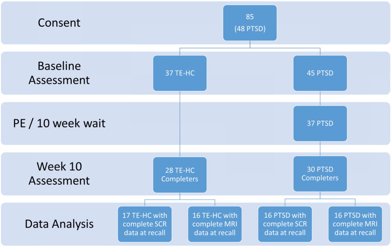
Fifty-eight participants (30 with PTSD, 28 trauma-exposed matched controls) underwent a 2-day behavioral fear conditioning, extinction, and recall paradigm during functional magnetic resonance imaging (fMRI). The same procedures were repeated 10 weeks later, after PTSD patients had completed prolonged exposure treatment. We analyzed fMRI data from 32 subjects (16 PTSD; 16 controls) and skin conductance response (SCR) data from 33 subjects (16 PTSD; 17 controls). Neural activity during extinction recall, SCR, and PTSD symptoms were compared across groups and over time.
PTSD patients exhibited pre- to post-treatment reduction in rostral anterior cingulate cortex (rACC) activation during extinction recall, and increase in functional coherence between the rACC and the ventromedial prefrontal cortex (vmPFC) and subgenual anterior cingulate cortex (sgACC). Reduced PTSD symptom severity from pre- to post-treatment was significantly associated with reduced subgenual ACC and parahippocampal activation during this task. SCR during the extinction recall phase did not significantly change with treatment in the PTSD group, but change in SCR was associated with reduction in PTSD symptom severity.
Prolonged exposure treatment appears to alter neural activation in PTSD patients during recall of fear extinction, and change in extinction recall (measured by SCR) is associated with symptom reduction. We discuss results in the context of neural systems involved in response to affective stimuli.
创伤后应激障碍(PTSD)的神经生物学模型表明,恐惧加工障碍在该疾病的维持中起作用。已证实存在消退回忆方面的特定缺陷,即对习得的消退的保持。虽然消退回忆缺陷以及前额叶和海马区的相关激活模式可将PTSD患者与对照组区分开来,但研究尚未考察治疗后的变化。我们研究了PTSD认知行为治疗前后消退回忆的行为和神经相关性。
58名参与者(30名PTSD患者,28名创伤暴露匹配对照组)在功能磁共振成像(fMRI)期间接受了为期2天的行为恐惧条件反射、消退和回忆范式。10周后,PTSD患者完成延长暴露治疗后,重复相同程序。我们分析了32名受试者(16名PTSD患者;16名对照组)的fMRI数据和33名受试者(16名PTSD患者;17名对照组)的皮肤电导反应(SCR)数据。比较了各组之间以及不同时间点的消退回忆期间的神经活动、SCR和PTSD症状。
PTSD患者在消退回忆期间,治疗前到治疗后,喙前扣带回皮质(rACC)激活减少,rACC与腹内侧前额叶皮质(vmPFC)和膝下前扣带回皮质(sgACC)之间的功能连贯性增加。治疗前到治疗后PTSD症状严重程度降低与该任务期间膝下ACC和海马旁回激活减少显著相关。PTSD组在消退回忆阶段的SCR随治疗无显著变化,但SCR的变化与PTSD症状严重程度降低相关。
延长暴露治疗似乎在PTSD患者恐惧消退回忆期间改变神经激活,并且消退回忆的变化(通过SCR测量)与症状减轻相关。我们在涉及对情感刺激反应的神经系统背景下讨论结果。