• 文献检索
  • 文档翻译
  • 深度研究
  • 学术资讯
  • Suppr Zotero 插件Zotero 插件
  • 邀请有礼
  • 套餐&价格
  • 历史记录
应用&插件
Suppr Zotero 插件Zotero 插件浏览器插件Mac 客户端Windows 客户端微信小程序
定价
高级版会员购买积分包购买API积分包
服务
文献检索文档翻译深度研究API 文档MCP 服务
关于我们
关于 Suppr公司介绍联系我们用户协议隐私条款
关注我们

Suppr 超能文献

核心技术专利:CN118964589B侵权必究
粤ICP备2023148730 号-1Suppr @ 2026

文献检索

告别复杂PubMed语法,用中文像聊天一样搜索,搜遍4000万医学文献。AI智能推荐,让科研检索更轻松。

立即免费搜索

文件翻译

保留排版,准确专业,支持PDF/Word/PPT等文件格式,支持 12+语言互译。

免费翻译文档

深度研究

AI帮你快速写综述,25分钟生成高质量综述,智能提取关键信息,辅助科研写作。

立即免费体验

γ-原钙黏蛋白结构多样性及其功能意义。

γ-Protocadherin structural diversity and functional implications.

机构信息

Department of Biochemistry and Molecular Biophysics, Columbia University, New York, United States.

Department of Systems Biology, Columbia University, New York, United States.

出版信息

Elife. 2016 Oct 26;5:e20930. doi: 10.7554/eLife.20930.

DOI:10.7554/eLife.20930
PMID:27782885
原文链接:https://pmc.ncbi.nlm.nih.gov/articles/PMC5106212/
Abstract

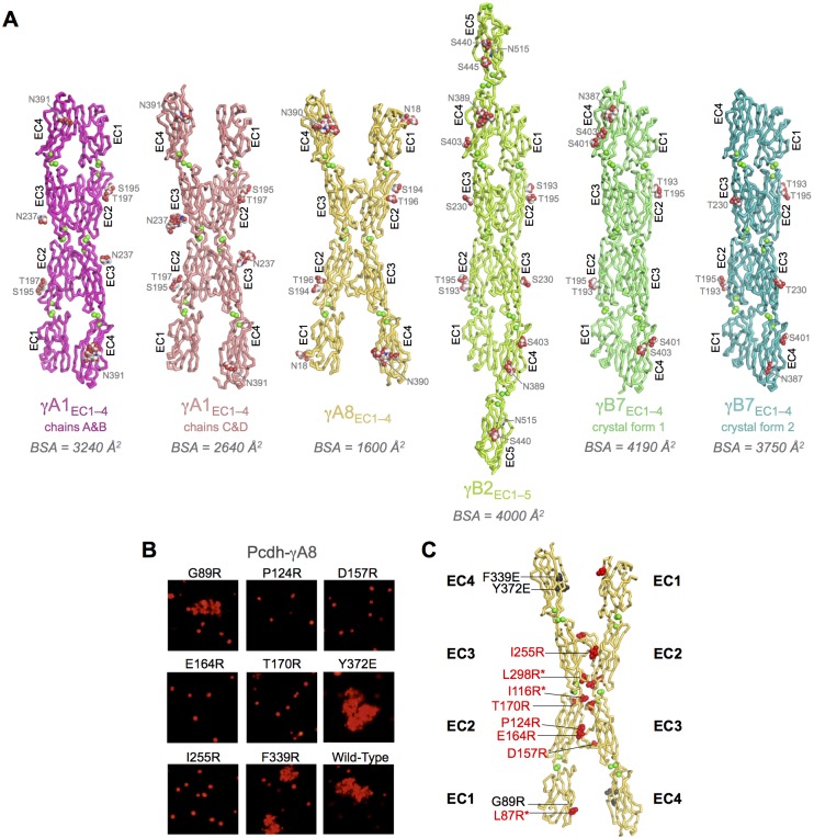
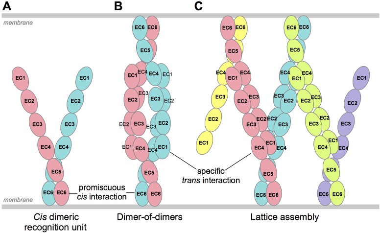
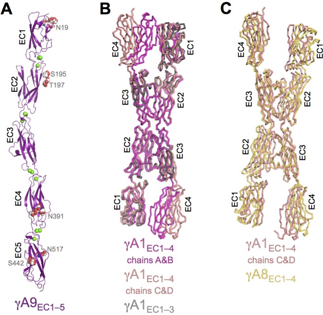
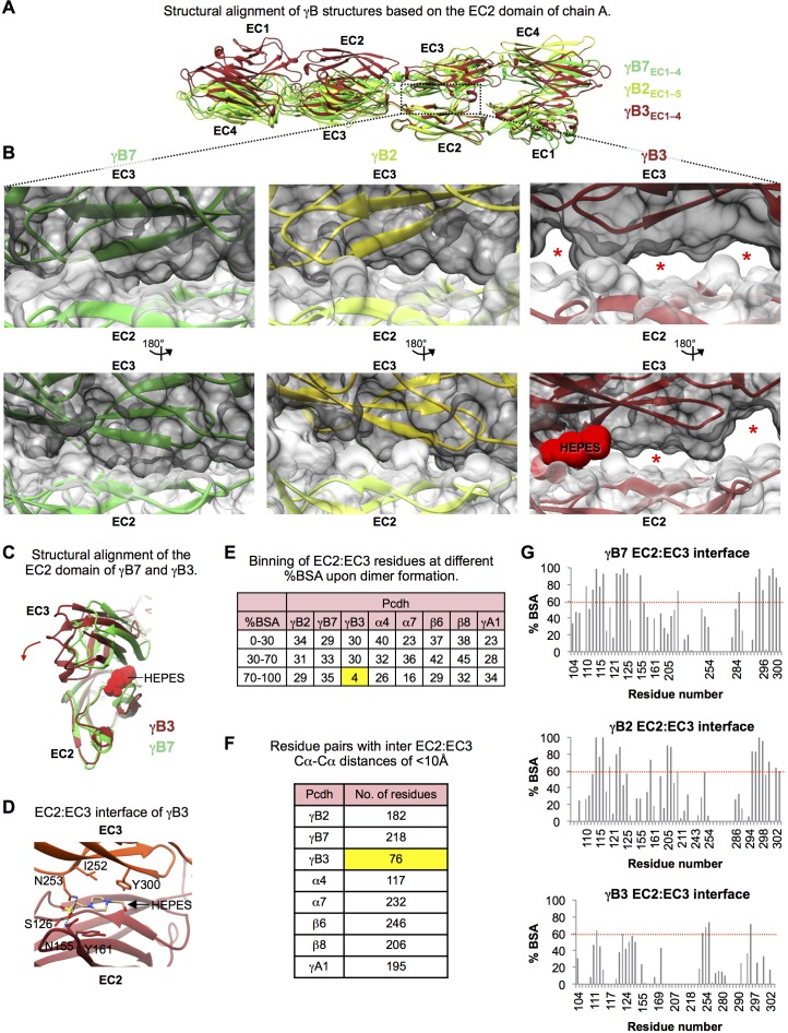
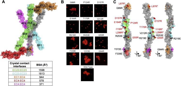
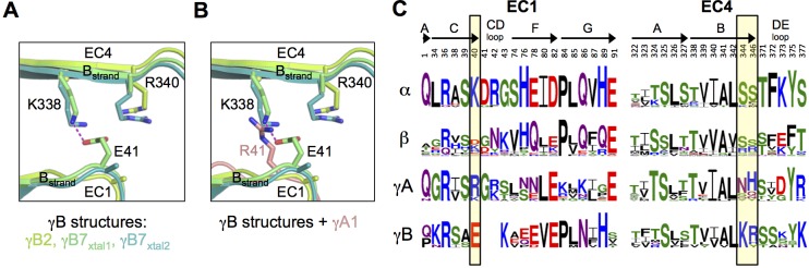
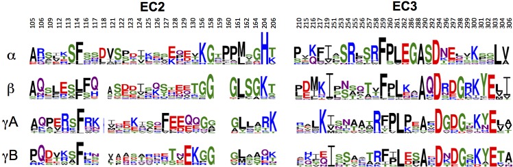
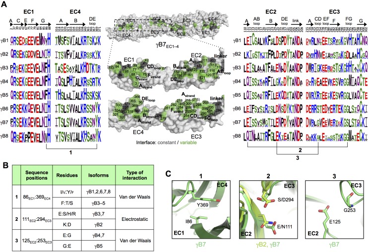
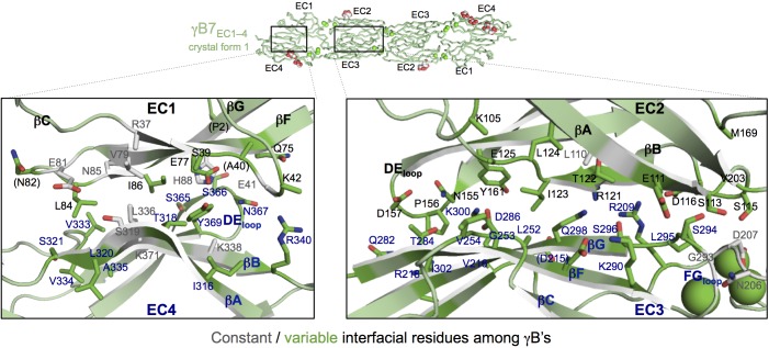
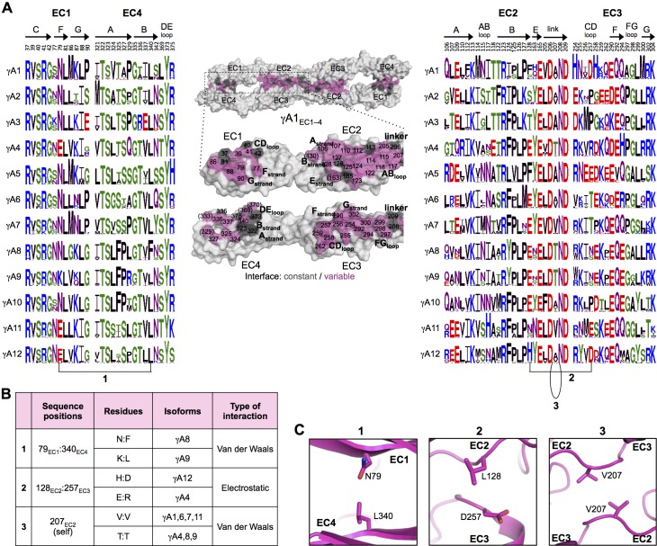
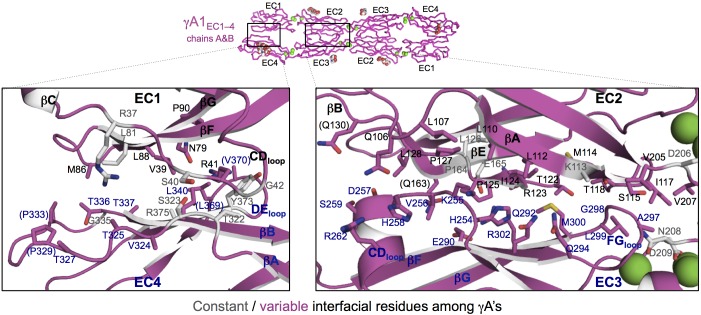
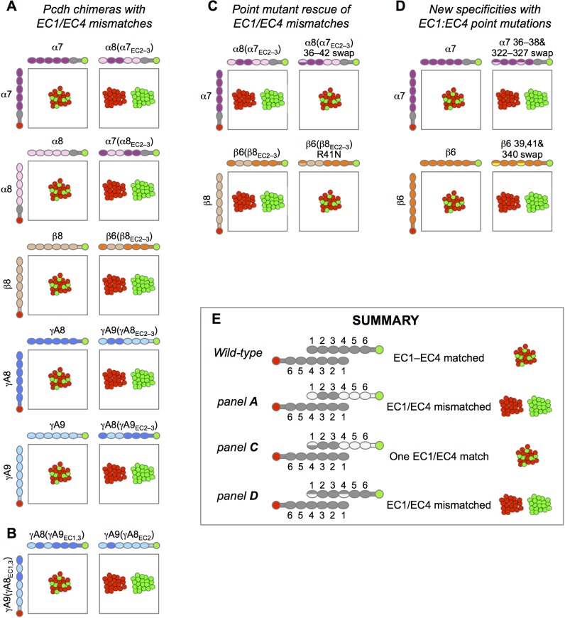
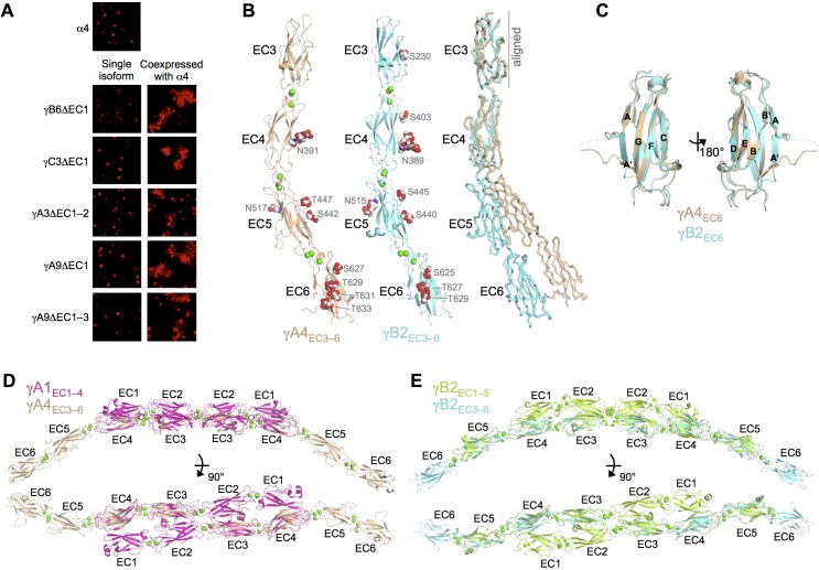
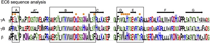
Stochastic cell-surface expression of α-, β-, and γ-clustered protocadherins (Pcdhs) provides vertebrate neurons with single-cell identities that underlie neuronal self-recognition. Here we report crystal structures of ectodomain fragments comprising cell-cell recognition regions of mouse γ-Pcdhs γA1, γA8, γB2, and γB7 revealing -homodimers, and of C-terminal ectodomain fragments from γ-Pcdhs γA4 and γB2, which depict -interacting regions in monomeric form. Together these structures span the entire γ-Pcdh ectodomain. The -dimer structures reveal determinants of γ-Pcdh isoform-specific homophilic recognition. We identified and structurally mapped -dimerization mutations to the C-terminal ectodomain structures. Biophysical studies showed that Pcdh ectodomains from γB-subfamily isoforms formed dimers, whereas γA isoforms did not, but both γA and γB isoforms could interact in with α-Pcdhs. Together, these data show how interaction specificity is distributed over all domains of the γ-Pcdh interface, and suggest that subfamily- or isoform-specific -interactions may play a role in the Pcdh-mediated neuronal self-recognition code.

摘要

随机的细胞表面α-、β-和γ-聚类原钙粘蛋白(Pcdh)的表达为脊椎动物神经元提供了单细胞身份,这是神经元自我识别的基础。在这里,我们报告了包含小鼠γ-Pcdh γA1、γA8、γB2 和 γB7 的细胞间识别区域的外域片段的晶体结构,揭示了同源二聚体,以及γ-Pcdh γA4 和 γB2 的 C 端外域片段,其以单体形式描绘了相互作用区域。这些结构共同跨越了整个γ-Pcdh 外域。-二聚体结构揭示了γ-Pcdh 同工型特异性同亲识别的决定因素。我们确定并在结构上对 C 端外域结构进行了 -二聚化突变。生物物理研究表明,γB 亚家族同工型的 Pcdh 外域形成二聚体,而 γA 同工型则不能,但 γA 和 γB 同工型都可以与α-Pcdh 相互作用。总之,这些数据表明了相互作用特异性是如何分布在γ-Pcdh 界面的所有结构域上的,并表明亚家族或同工型特异性 -相互作用可能在 Pcdh 介导的神经元自我识别代码中发挥作用。

https://cdn.ncbi.nlm.nih.gov/pmc/blobs/0a79/5106212/befc5387b138/elife-20930-fig7.jpg
https://cdn.ncbi.nlm.nih.gov/pmc/blobs/0a79/5106212/13ee1b4f3265/elife-20930-fig1.jpg
https://cdn.ncbi.nlm.nih.gov/pmc/blobs/0a79/5106212/4bc187dc5966/elife-20930-fig1-figsupp1.jpg
https://cdn.ncbi.nlm.nih.gov/pmc/blobs/0a79/5106212/f9eaf0b96624/elife-20930-fig1-figsupp2.jpg
https://cdn.ncbi.nlm.nih.gov/pmc/blobs/0a79/5106212/272c179b7746/elife-20930-fig1-figsupp3.jpg
https://cdn.ncbi.nlm.nih.gov/pmc/blobs/0a79/5106212/61e51ea67590/elife-20930-fig1-figsupp4.jpg
https://cdn.ncbi.nlm.nih.gov/pmc/blobs/0a79/5106212/85bdbccef411/elife-20930-fig2.jpg
https://cdn.ncbi.nlm.nih.gov/pmc/blobs/0a79/5106212/1b411efd8170/elife-20930-fig2-figsupp1.jpg
https://cdn.ncbi.nlm.nih.gov/pmc/blobs/0a79/5106212/0ea47be669bf/elife-20930-fig3.jpg
https://cdn.ncbi.nlm.nih.gov/pmc/blobs/0a79/5106212/72f510a18e16/elife-20930-fig3-figsupp1.jpg
https://cdn.ncbi.nlm.nih.gov/pmc/blobs/0a79/5106212/7aa5a349beaa/elife-20930-fig3-figsupp2.jpg
https://cdn.ncbi.nlm.nih.gov/pmc/blobs/0a79/5106212/5c599b32b802/elife-20930-fig4.jpg
https://cdn.ncbi.nlm.nih.gov/pmc/blobs/0a79/5106212/bd1d4a391acc/elife-20930-fig4-figsupp1.jpg
https://cdn.ncbi.nlm.nih.gov/pmc/blobs/0a79/5106212/5d0fd11173dd/elife-20930-fig4-figsupp2.jpg
https://cdn.ncbi.nlm.nih.gov/pmc/blobs/0a79/5106212/4d7fdbca0308/elife-20930-fig5.jpg
https://cdn.ncbi.nlm.nih.gov/pmc/blobs/0a79/5106212/a21fcd722ecc/elife-20930-fig5-figsupp1.jpg
https://cdn.ncbi.nlm.nih.gov/pmc/blobs/0a79/5106212/ab5ae165c15d/elife-20930-fig6.jpg
https://cdn.ncbi.nlm.nih.gov/pmc/blobs/0a79/5106212/eba819943796/elife-20930-fig6-figsupp1.jpg
https://cdn.ncbi.nlm.nih.gov/pmc/blobs/0a79/5106212/ca8c1d08995d/elife-20930-fig6-figsupp2.jpg
https://cdn.ncbi.nlm.nih.gov/pmc/blobs/0a79/5106212/befc5387b138/elife-20930-fig7.jpg

相似文献

1
γ-Protocadherin structural diversity and functional implications.γ-原钙黏蛋白结构多样性及其功能意义。
Elife. 2016 Oct 26;5:e20930. doi: 10.7554/eLife.20930.
2
Protocadherin -dimer architecture and recognition unit diversity.原钙黏蛋白二聚体结构与识别单位多样性。
Proc Natl Acad Sci U S A. 2017 Nov 14;114(46):E9829-E9837. doi: 10.1073/pnas.1713449114. Epub 2017 Oct 30.
3
Structural Basis of Diverse Homophilic Recognition by Clustered α- and β-Protocadherins.成簇的α-和β-原钙黏蛋白介导多种同嗜性识别的结构基础
Neuron. 2016 May 18;90(4):709-23. doi: 10.1016/j.neuron.2016.04.004. Epub 2016 May 5.
4
Visualization of clustered protocadherin neuronal self-recognition complexes.聚集原钙黏蛋白神经元自我识别复合物的可视化。
Nature. 2019 May;569(7755):280-283. doi: 10.1038/s41586-019-1089-3. Epub 2019 Apr 10.
5
Combinatorial homophilic interaction between gamma-protocadherin multimers greatly expands the molecular diversity of cell adhesion.γ-原钙黏蛋白多聚体的组合同性相互作用极大地扩展了细胞黏附的分子多样性。
Proc Natl Acad Sci U S A. 2010 Aug 17;107(33):14893-8. doi: 10.1073/pnas.1004526107. Epub 2010 Aug 2.
6
Antiparallel protocadherin homodimers use distinct affinity- and specificity-mediating regions in cadherin repeats 1-4.反向平行原钙黏蛋白同型二聚体在钙黏蛋白重复序列1-4中使用不同的亲和力和特异性介导区域。
Elife. 2016 Jul 29;5:e18449. doi: 10.7554/eLife.18449.
7
Single-cell identity generated by combinatorial homophilic interactions between α, β, and γ protocadherins.由α、β和γ原钙黏蛋白之间的组合性嗜同性相互作用产生的单细胞身份。
Cell. 2014 Aug 28;158(5):1045-1059. doi: 10.1016/j.cell.2014.07.012.
8
Differential expression of individual gamma-protocadherins during mouse brain development.小鼠脑发育过程中单个γ-原钙黏蛋白的差异表达。
Mol Cell Neurosci. 2005 Aug;29(4):603-16. doi: 10.1016/j.mcn.2005.05.001.
9
Structure and Sequence Analyses of Clustered Protocadherins Reveal Antiparallel Interactions that Mediate Homophilic Specificity.成簇原钙黏蛋白的结构与序列分析揭示了介导嗜同性特异性的反平行相互作用。
Structure. 2015 Nov 3;23(11):2087-98. doi: 10.1016/j.str.2015.09.005. Epub 2015 Oct 15.
10
How clustered protocadherin binding specificity is tuned for neuronal self-/nonself-recognition.成簇原钙黏蛋白的结合特异性是如何针对神经元的自我/非自我识别进行调节的。
Elife. 2022 Mar 7;11:e72416. doi: 10.7554/eLife.72416.

引用本文的文献

1
Surface delivery quantification reveals distinct trafficking efficiencies among clustered protocadherin isoforms.表面递送定量分析揭示了成簇原钙黏蛋白异构体之间不同的转运效率。
Proc Natl Acad Sci U S A. 2025 Aug 5;122(31):e2514178122. doi: 10.1073/pnas.2514178122. Epub 2025 Jul 30.
2
Surface delivery quantification reveals distinct trafficking efficiencies among clustered protocadherin isoforms.表面递送定量分析揭示了成簇原钙黏蛋白亚型之间不同的运输效率。
bioRxiv. 2025 Jun 6:2024.09.23.614616. doi: 10.1101/2024.09.23.614616.
3
Diversity of clustered protocadherin-α genes in neuronal identity and its role in short-term specific associative memory formation.

本文引用的文献

1
Antiparallel protocadherin homodimers use distinct affinity- and specificity-mediating regions in cadherin repeats 1-4.反向平行原钙黏蛋白同型二聚体在钙黏蛋白重复序列1-4中使用不同的亲和力和特异性介导区域。
Elife. 2016 Jul 29;5:e18449. doi: 10.7554/eLife.18449.
2
Structural basis of adhesive binding by desmocollins and desmogleins.桥粒芯蛋白和桥粒芯胶蛋白进行黏附结合的结构基础。
Proc Natl Acad Sci U S A. 2016 Jun 28;113(26):7160-5. doi: 10.1073/pnas.1606272113. Epub 2016 Jun 13.
3
Structural Basis of Diverse Homophilic Recognition by Clustered α- and β-Protocadherins.
成簇原钙黏蛋白-α基因在神经元身份中的多样性及其在短期特异性联想记忆形成中的作用。
Sci Rep. 2025 Jun 2;15(1):19334. doi: 10.1038/s41598-025-02546-2.
4
Structural insights into the in situ assembly of clustered protocadherin γB4.成簇原钙黏蛋白γB4原位组装的结构见解
Nat Commun. 2025 Feb 16;16(1):1682. doi: 10.1038/s41467-025-56948-x.
5
Computational investigation of missense somatic mutations in cancer and potential links to pH-dependence and proteostasis.计算研究癌症中的错义体细胞突变及其与 pH 依赖性和蛋白质平衡的潜在联系。
PLoS One. 2024 Nov 19;19(11):e0314022. doi: 10.1371/journal.pone.0314022. eCollection 2024.
6
Clustered protocadherin -interactions are required for combinatorial cell-cell recognition underlying neuronal self-avoidance.簇集原钙黏蛋白相互作用对于神经元自我回避所必需的组合细胞间识别。
Proc Natl Acad Sci U S A. 2024 Jul 16;121(29):e2319829121. doi: 10.1073/pnas.2319829121. Epub 2024 Jul 8.
7
Ubiquitination of the protocadherin-γA3 variable cytoplasmic domain modulates cell-cell interaction.原钙黏蛋白γA3可变胞质结构域的泛素化调节细胞间相互作用。
Front Cell Dev Biol. 2023 Sep 18;11:1261048. doi: 10.3389/fcell.2023.1261048. eCollection 2023.
8
Visualization of homophilic interaction of clustered protocadherin in neurons.神经元中同源聚集原钙黏蛋白相互作用的可视化。
Proc Natl Acad Sci U S A. 2023 Sep 19;120(38):e2301003120. doi: 10.1073/pnas.2301003120. Epub 2023 Sep 11.
9
γ-Protocadherins control synapse formation and peripheral branching of touch sensory neurons.γ-原钙黏蛋白控制着突触的形成和触觉感觉神经元的外围分支。
Neuron. 2023 Jun 7;111(11):1776-1794.e10. doi: 10.1016/j.neuron.2023.03.012. Epub 2023 Apr 6.
10
Structure of the planar cell polarity cadherins Fat4 and Dachsous1.平面细胞极性钙黏蛋白 Fat4 和 Dachsous1 的结构。
Nat Commun. 2023 Feb 16;14(1):891. doi: 10.1038/s41467-023-36435-x.
成簇的α-和β-原钙黏蛋白介导多种同嗜性识别的结构基础
Neuron. 2016 May 18;90(4):709-23. doi: 10.1016/j.neuron.2016.04.004. Epub 2016 May 5.
4
Homophilic Protocadherin Cell-Cell Interactions Promote Dendrite Complexity.同源性原钙黏蛋白细胞间相互作用促进树突复杂性。
Cell Rep. 2016 May 3;15(5):1037-1050. doi: 10.1016/j.celrep.2016.03.093. Epub 2016 Apr 21.
5
Structure and Sequence Analyses of Clustered Protocadherins Reveal Antiparallel Interactions that Mediate Homophilic Specificity.成簇原钙黏蛋白的结构与序列分析揭示了介导嗜同性特异性的反平行相互作用。
Structure. 2015 Nov 3;23(11):2087-98. doi: 10.1016/j.str.2015.09.005. Epub 2015 Oct 15.
6
Molecular logic of neuronal self-recognition through protocadherin domain interactions.通过原钙黏蛋白结构域相互作用实现神经元自我识别的分子逻辑
Cell. 2015 Oct 22;163(3):629-42. doi: 10.1016/j.cell.2015.09.026. Epub 2015 Oct 17.
7
A hybrid method for protein-protein interface prediction.一种用于蛋白质-蛋白质界面预测的混合方法。
Protein Sci. 2016 Jan;25(1):159-65. doi: 10.1002/pro.2744. Epub 2015 Jul 21.
8
Protocadherin-dependent dendritic self-avoidance regulates neural connectivity and circuit function.原钙黏蛋白依赖性树突自我回避调节神经连接和回路功能。
Elife. 2015 Jul 3;4:e08964. doi: 10.7554/eLife.08964.
9
Single-cell identity generated by combinatorial homophilic interactions between α, β, and γ protocadherins.由α、β和γ原钙黏蛋白之间的组合性嗜同性相互作用产生的单细胞身份。
Cell. 2014 Aug 28;158(5):1045-1059. doi: 10.1016/j.cell.2014.07.012.
10
Probabilistic splicing of Dscam1 establishes identity at the level of single neurons.Dscam1 的概率剪接在单个神经元水平上建立了身份。
Cell. 2013 Nov 21;155(5):1166-77. doi: 10.1016/j.cell.2013.10.018.