Evin Morgane, Broadhouse Kathryn M, Callaghan Fraser M, McGrath Rachel T, Glastras Sarah, Kozor Rebecca, Hocking Samantha L, Lamy Jérôme, Redheuil Alban, Kachenoura Nadjia, Fulcher Greg R, Figtree Gemma A, Grieve Stuart M
Sydney Translational Imaging Laboratory, Heart Research Institute, Charles Perkins Centre, University of Sydney, Camperdown, NSW, 2006, Australia.
Sydney Medical School, University of Sydney, Camperdown, Australia.
Cardiovasc Diabetol. 2016 Dec 22;15(1):164. doi: 10.1186/s12933-016-0481-7.
Diastolic dysfunction is a major cause of morbidity in obese individuals. We aimed to assess the ability of magnetic resonance imaging (MRI) derived left atrial (LA) strain to detect early diastolic dysfunction in individuals with obesity and type 2 diabetes, and to explore the association between cardiac adipose tissue and LA function.
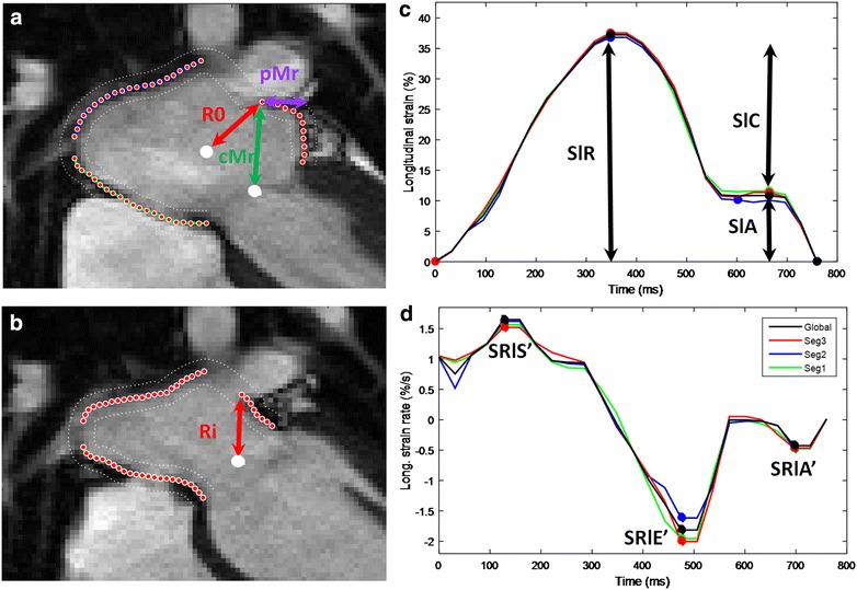
Twenty patients with obesity and T2D (55 ± 8 years) and nineteen healthy controls (48 ± 13 years) were imaged using cine steady state free precession and 2-point Dixon cardiovascular magnetic resonance. LA function was quantified using a feature tracking technique with definition of phasic longitudinal strain and strain rates, as well as radial motion fraction and radial velocities.
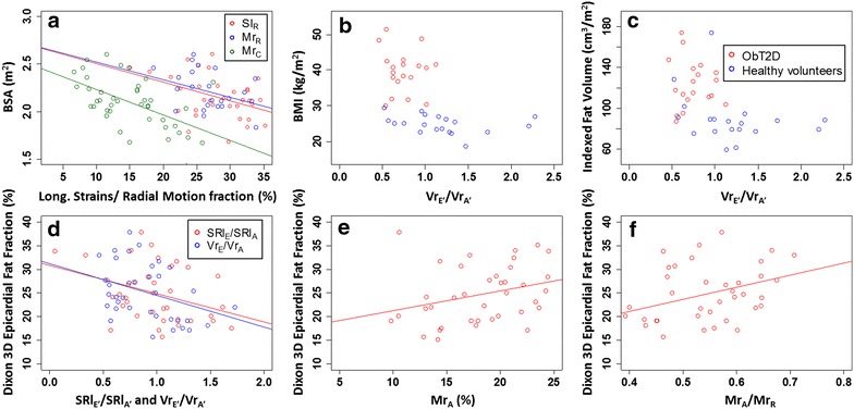
Systolic left ventricular size and function were similar between the obesity and type 2 diabetes and control groups by MRI. All patients except four had normal diastolic assessment by echocardiography. In contrast, measures of LA function using magnetic resonance feature tracking were uniformly altered in the obesity and type 2 diabetes group only. Although there was no significant difference in intra-myocardial fat fraction, Dixon 3D epicardial fat volume(EFV) was significantly elevated in the obesity and type 2 diabetes versus control group (135 ± 31 vs. 90 ± 30 mL/m, p < 0.001). There were significant correlations between LA functional indices and both BMI and EFV (p ≤ 0.007).
LA MRI-strain may be a sensitive tool for the detection of early diastolic dysfunction in individuals with obesity and type 2 diabetes and correlated with BMI and epicardial fat supporting a possible association between adiposity and LA strain. Trials Registration Australian New Zealand Clinical Trials Registry No. ACTRN12613001069741.
舒张功能障碍是肥胖个体发病的主要原因。我们旨在评估磁共振成像(MRI)衍生的左心房(LA)应变检测肥胖和2型糖尿病个体早期舒张功能障碍的能力,并探讨心脏脂肪组织与LA功能之间的关联。
对20例肥胖和2型糖尿病患者(55±8岁)和19例健康对照者(48±13岁)进行电影稳态自由进动和两点Dixon心血管磁共振成像。使用特征跟踪技术对LA功能进行量化,定义阶段性纵向应变和应变率,以及径向运动分数和径向速度。
通过MRI,肥胖和2型糖尿病组与对照组之间的收缩期左心室大小和功能相似。除4例患者外,所有患者经超声心动图评估舒张功能均正常。相比之下,仅肥胖和2型糖尿病组使用磁共振特征跟踪的LA功能测量值均发生了改变。尽管心肌内脂肪分数无显著差异,但肥胖和2型糖尿病组的Dixon 3D心外膜脂肪体积(EFV)显著高于对照组(135±31 vs. 90±30 mL/m,p<0.001)。LA功能指标与BMI和EFV均存在显著相关性(p≤0.007)。
LA MRI应变可能是检测肥胖和2型糖尿病个体早期舒张功能障碍的敏感工具,且与BMI和心外膜脂肪相关,支持肥胖与LA应变之间可能存在关联。试验注册澳大利亚新西兰临床试验注册中心编号ACTRN12613001069741。