Braat Manon N G J A, de Jong Hugo W, Seinstra Beatrijs A, Scholten Mike V, van den Bosch Maurice A A J, Lam Marnix G E H
Department of Radiology and Nuclear Medicine, University Medical Center Utrecht, Heidelberglaan 100, 3584 CX, Utrecht, The Netherlands.
Department of Radiology and Nuclear Medicine, Meander Medical Center, Amersfoort, The Netherlands.
EJNMMI Res. 2017 Dec;7(1):2. doi: 10.1186/s13550-016-0248-x. Epub 2017 Jan 5.
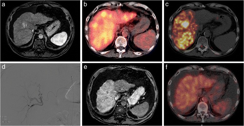
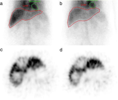
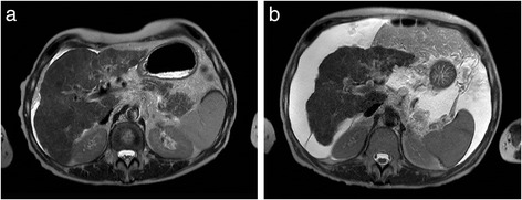
Routine work-up for transarterial radioembolization, based on clinical and laboratory parameters, sometimes fails, resulting in severe hepatotoxicity in up to 5% of patients. Quantitative assessment of the pretreatment liver function and its segmental distribution, using hepatobiliary scintigraphy may improve patient selection and treatment planning. A case series will be presented to illustrate the potential of this technique. Hepatocellular carcinoma patients with cirrhosis (Child-Pugh A and B) underwent hepatobiliary scintigraphy pre- and 3 months post-radioembolization as part of a prospective study protocol, which was prematurely terminated because of limited accrual. Included patients were analysed together with their clinical, laboratory and treatment data.
Pretreatment-corrected Tc-mebrofenin liver uptake rates were marginal (1.8-3.0%/min/m), despite acceptable clinical and laboratory parameters. Posttreatment liver functions seriously declined (corrected Tc-mebrofenin liver uptake rates: 0.6-2.4%/min/m), resulting in lethal radioembolization-induced liver disease in two out of three patients.
Hepatobiliary scintigraphy may be of added value during work-up for radioembolization, to estimate liver function reserve and its segmental distribution, especially in patients with underlying cirrhosis, for whom analysis of clinical and laboratory parameters may not be sufficient.
基于临床和实验室参数的经动脉放射性栓塞常规检查有时会失败,导致高达5%的患者出现严重肝毒性。使用肝胆闪烁显像对治疗前肝功能及其节段分布进行定量评估,可能会改善患者选择和治疗规划。本文将通过一个病例系列来说明该技术的潜力。作为一项前瞻性研究方案的一部分,肝硬化(Child-Pugh A级和B级)的肝细胞癌患者在放射性栓塞术前和术后3个月接受了肝胆闪烁显像检查,该研究方案因入组人数有限而提前终止。对纳入的患者及其临床、实验室和治疗数据进行了分析。
尽管临床和实验室参数尚可,但治疗前校正后的锝-美罗芬宁肝脏摄取率较低(1.8 - 3.0%/分钟/米)。治疗后肝功能严重下降(校正后的锝-美罗芬宁肝脏摄取率:0.6 - 2.4%/分钟/米),导致三名患者中有两名死于放射性栓塞诱发的肝病。
在放射性栓塞检查过程中,肝胆闪烁显像可能具有附加价值,可用于评估肝功能储备及其节段分布,特别是对于潜在肝硬化患者,临床和实验室参数分析可能并不充分。