Liu Jianghong, Liang Peipeng, Yin Linlin, Shu Ni, Zhao Tengda, Xing Yi, Li Fangyu, Zhao Zhilian, Li Kuncheng, Han Ying
Department of Neurology, Xuan Wu Hospital, Capital Medical University, Beijing, China.
Department of Radiology, Xuan Wu Hospital, Capital Medical University, Beijing, China.
PLoS One. 2017 Jan 20;12(1):e0170185. doi: 10.1371/journal.pone.0170185. eCollection 2017.
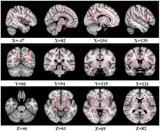
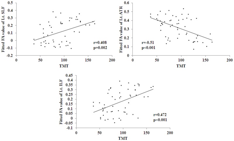
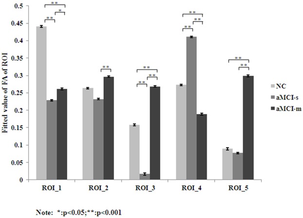
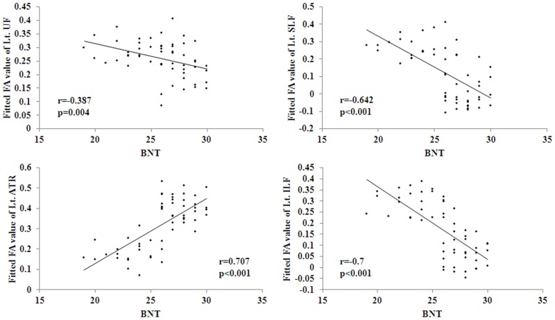
White matter (WM) degeneration has been found during the course of cognitive decline in both Alzheimer's disease (AD) and amnestic mild cognitive impairment (aMCI), however, it is unclear whether there are different WM microstructural abnormalities between two subtypes of aMCI, including single domain aMCI (aMCI-s) and multiple domain aMCI (aMCI-m). Thirty-two patients of aMCI single-domain (aMCI-s), twenty-three patients of aMCI multiple-domain (aMCI-m) and twenty-three healthy normal controls (NC) participated in this study. Neuropsychological measures and diffusion tensor imaging (DTI) data were acquired from each subject and tract-based spatial statistics (TBSS) was implemented. It was found that both aMCI groups showed significantly reduced fractional anisotropy (FA) in the right superior longitudinal fasciculus (SLF) than NC. It was also identified that, as compared to aMCI-m, aMCI-s showed significantly decreased FA in the left SLF, left uncinate fasciculus (UF) and left inferior longitudinal fasciculus (ILF), while significantly increased FA in the left anterior thalamic radiation (ATR). The correlation analysis showed that FA values in the regions with group difference were significantly correlated with cognitive functions as measured by Boston naming test and trail making test. These results suggested that the variations of aMCI may be differentiated by FA indexes and DTI may help to understand why specific signs and symptoms occur in patients.
在阿尔茨海默病(AD)和遗忘型轻度认知障碍(aMCI)的认知衰退过程中均发现了白质(WM)变性,然而,尚不清楚aMCI的两种亚型,即单领域aMCI(aMCI-s)和多领域aMCI(aMCI-m)之间是否存在不同的WM微观结构异常。32名单领域aMCI(aMCI-s)患者、23名多领域aMCI(aMCI-m)患者和23名健康正常对照(NC)参与了本研究。从每个受试者获取神经心理学测量和扩散张量成像(DTI)数据,并实施基于束的空间统计学(TBSS)。结果发现,与NC相比,两个aMCI组右侧上纵束(SLF)的分数各向异性(FA)均显著降低。还发现,与aMCI-m相比,aMCI-s左侧SLF、左侧钩束(UF)和左侧下纵束(ILF)的FA显著降低,而左侧丘脑前辐射(ATR)的FA显著增加。相关性分析表明,具有组间差异区域的FA值与波士顿命名测试和连线测试所测量的认知功能显著相关。这些结果表明,aMCI的变异可能通过FA指数进行区分,DTI可能有助于理解患者为何会出现特定的体征和症状。