Calabrese Cecilia, Gilli Marina, De Rosa Nicolina, Di Crescenzo Vincenzo, Zeppa Pio, Vitale Carolina, Vatrella Alessandro
Department of Cardio-Thoracic and Respiratory Sciences, Second University of Naples, Naples, Italy.
AORN Ospedale dei Colli, Division of Pulmonary Oncology, Naples, Italy.
Open Med (Wars). 2016 Jun 23;11(1):158-162. doi: 10.1515/med-2016-0025. eCollection 2016.
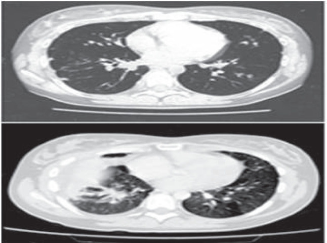
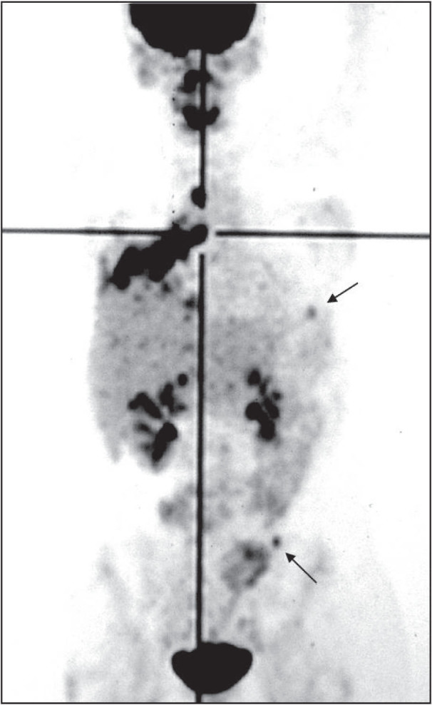
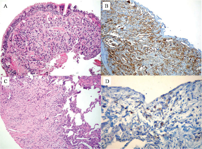
In this report we describe a case of pulmonary epithelioid hemangioendothelioma (PEH) in a young woman. The neoplasm manifested with dry cough, chest pain, finger clubbing, and multiple bilateral pulmonary nodules on chest x-ray and computed tomographic (CT) scan. She underwent thoracoscopy, and the histological features of the lung biopsies were initially interpreted as consistent with a not-well-defined interstitial lung disease. Our patient was clinically and radiologically stable over a period of four years, after which the disease progressed to involve not only the lung but also mediastinal lymph nodes, liver and bone. Fiberoptic bronchoscopy showed subtotal occlusion of the right middle and lower lobe bronchi. The histologic examination of bronchial biopsies revealed a poorly differentiated neoplasm immunohistochemically positive for vimentin and vascular markers CD31, CD34 and Factor VIII. A diagnosis of malignant hemangioendothelioma was made. Positron emission tomography (PET) is more sensitive than CT scan and bone scintigraphy in detecting PEH metastases. Furthermore, 18-fluorodeoxyglucose (FDG) uptake seems to be related to the grade of malignancy of PEH lesions. Therefore, we suggest that FDG-PET should be included in the staging system and follow-up of PEH.
在本报告中,我们描述了一名年轻女性患肺上皮样血管内皮瘤(PEH)的病例。该肿瘤表现为干咳、胸痛、杵状指,胸部X线和计算机断层扫描(CT)显示双侧肺部有多个结节。她接受了胸腔镜检查,肺活检的组织学特征最初被解释为与一种界限不清的间质性肺病相符。我们的患者在四年时间里临床和放射学表现稳定,此后病情进展,不仅累及肺部,还累及纵隔淋巴结、肝脏和骨骼。纤维支气管镜检查显示右中叶和下叶支气管部分阻塞。支气管活检的组织学检查显示为低分化肿瘤,免疫组化显示波形蛋白及血管标志物CD31、CD34和因子VIII呈阳性。诊断为恶性血管内皮瘤。正电子发射断层扫描(PET)在检测PEH转移方面比CT扫描和骨闪烁显像更敏感。此外,18-氟脱氧葡萄糖(FDG)摄取似乎与PEH病变的恶性程度有关。因此,我们建议FDG-PET应纳入PEH的分期系统和随访中。