Department of Medical Radiology, University Hospital Zurich.
Department of Radiology, Nippon Medical School.
Magn Reson Med Sci. 2018 Jan 10;17(1):13-20. doi: 10.2463/mrms.mp.2016-0074. Epub 2017 Mar 27.
To assess the clinical feasibility of time-resolved 3D phase contrast (4D Flow) MRI assessment of the ophthalmic artery (OphA) flow in patients with internal carotid artery stenosis (ICS).
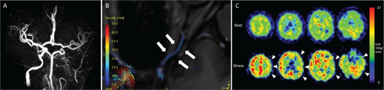
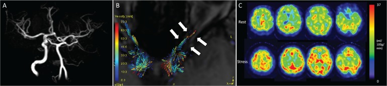
Twenty-one consecutive patients with unilateral ICS were recruited. 4D Flow MRI and acetazolamide-stress brain perfusion single photon emission computed tomography (SPECT) were performed. The flow direction on the affected-side OphA was categorized into native flow (anterograde or unclear) and non-native flow (retrograde flow) based on 4D Flow MRI. In the affected-side middle cerebral artery (MCA) territory, the ratio of rest cerebral blood flow to normal control (RCBF) and cerebral vascular reserve (CVR) were calculated from SPECT dataset. High-risk patients were defined based on the previous large cohort study (RCBF < 80% and CVRMCA < 10%).
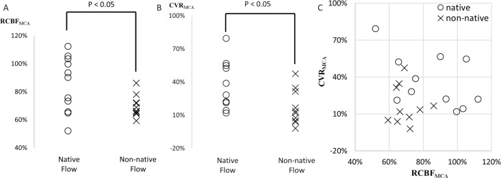
Eleven patients had native OphA flow (4 anterograde, 7 unclear) and the remaining 10 had non-native OphA flow. RCBF and CVR each were significantly lower in non-native flow group (84.9 ± 18.9% vs. 69.8 ± 7.3%, P < 0.05; 36.4 ± 20.6% vs. 17.0 ± 15.0%, P < 0.05). Four patients in the non-native flow group and none in the native flow group were confirmed as high-risk (Sensitivity/Specificity, 1.00/0.65).
The 6 min standard 4D Flow MRI assessment of OphA in patients with ICS can predict intracranial hemodynamic impairment.
评估时间分辨 3D 相位对比(4D Flow)MRI 评估颈内动脉狭窄(ICS)患者眼动脉(OphA)血流的临床可行性。
连续招募了 21 例单侧 ICS 患者。进行 4D Flow MRI 和乙酰唑胺应激脑灌注单光子发射计算机断层扫描(SPECT)。根据 4D Flow MRI 将患侧 OphA 的血流方向分为原生血流(顺行或不清)和非原生血流(逆行血流)。在患侧大脑中动脉(MCA)区域,从 SPECT 数据集计算静息脑血流与正常对照的比值(RCBF)和脑血流储备(CVR)。根据之前的大队列研究,将高危患者定义为 RCBF < 80%和 CVRMCA < 10%。
11 例患者 OphA 血流为原生(4 例顺行,7 例不清),其余 10 例为非原生血流。非原生血流组 RCBF 和 CVR 均显著降低(84.9 ± 18.9% vs. 69.8 ± 7.3%,P < 0.05;36.4 ± 20.6% vs. 17.0 ± 15.0%,P < 0.05)。非原生血流组有 4 例患者被确认为高危(敏感性/特异性,1.00/0.65),而原生血流组无患者为高危。
6 分钟标准 4D Flow MRI 评估 ICS 患者的 OphA 可以预测颅内血流动力学受损。