Repp Felix, Kollmannsberger Philip, Roschger Andreas, Kerschnitzki Michael, Berzlanovich Andrea, Gruber Gerlinde M, Roschger Paul, Wagermaier Wolfgang, Weinkamer Richard
Max Planck Institute of Colloids and Interfaces, Department of Biomaterials, D-14424 Potsdam, Germany.
Max Planck Institute of Colloids and Interfaces, Department of Biomaterials, D-14424 Potsdam, Germany; ETH Zürich, Laboratory of Applied Mechanobiology, Department of Health Sciences and Technology, CH-8093 Zurich, Switzerland.
Bone Rep. 2017 Mar 15;6:101-108. doi: 10.1016/j.bonr.2017.03.001. eCollection 2017 Jun.
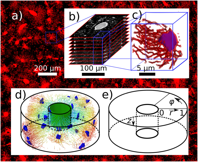
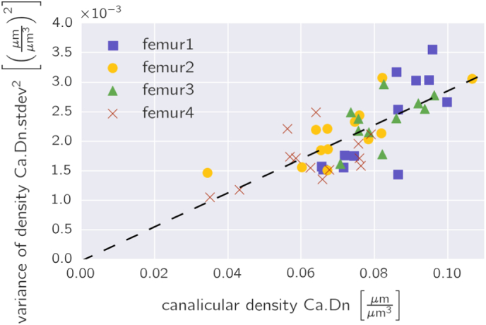
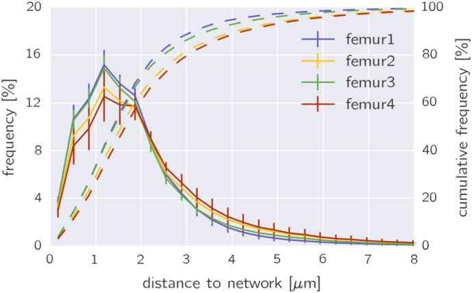
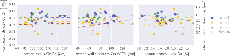
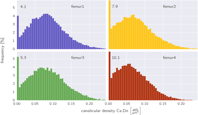
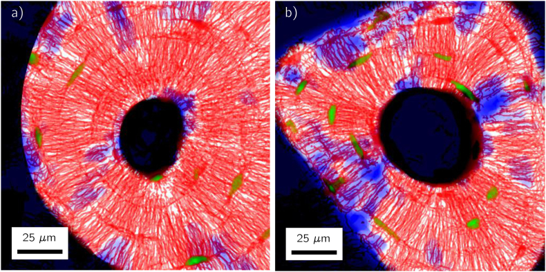
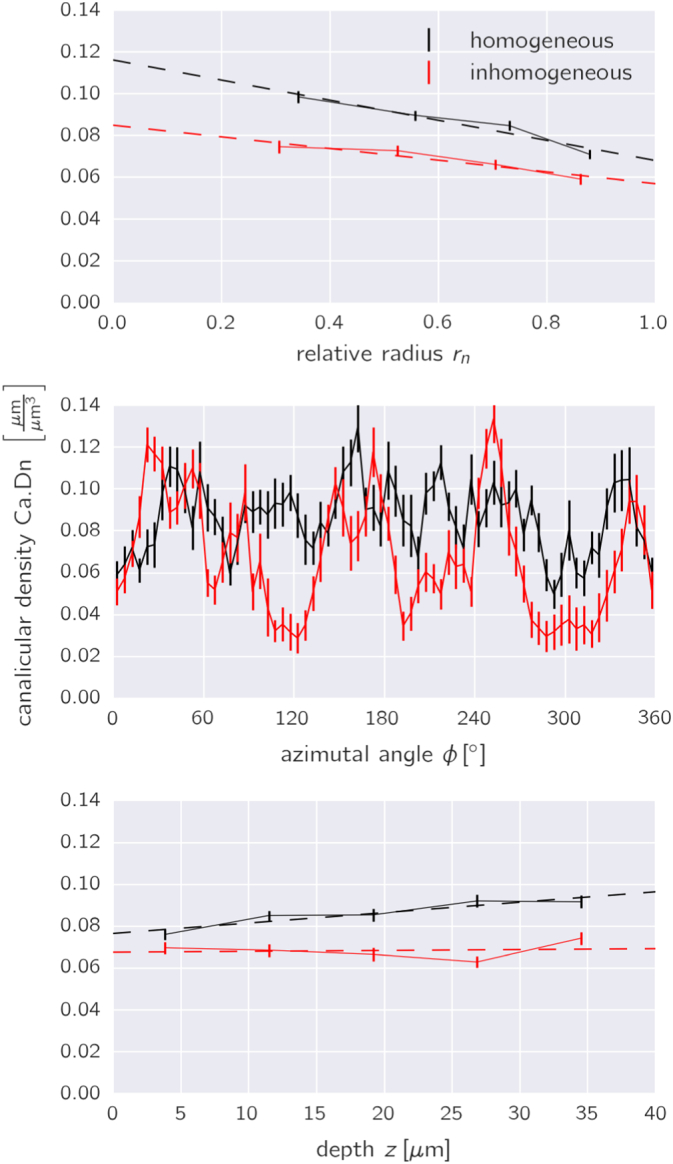
Osteocytes interconnect with each other forming an intricate cell network within the mineralized bone matrix. One important function of the osteocyte network is the mechano-regulation of bone remodeling, where a possible mechanism includes the fluid flow through the porosity housing the cell network - the osteocyte lacuno-canalicular network (OLCN). In our study the OLCN in human osteons was three-dimensionally imaged with the aim to obtain a quantitative description of the canalicular density and spatial variations of this quantity within osteons. The topology of the OLCN was determined by first staining the bone samples with rhodamine, then imaging the OLCN with confocal laser scanning microscopy and finally using image analysis to obtain a skeletonized version of the network for further analysis. In total 49 osteons were studied from the femoral cortical bone of four different middle-aged healthy women. The mean canalicular density given as length of the canaliculi in a unit volume was 0.074 ± 0.015 μm/μm (corresponding to 74 km/cm). No correlation was found between the canalicular density and neither the size of the osteon nor the volume fraction occupied by osteocyte lacunae. Within osteons the canalicular density varied substantially with larger regions without any network. On average the canalicular density decreases when moving from the Haversian canal outwards towards the cement line. We hypothesize that a decrease in accessible canaliculi with tissue age as a result of micropetrosis can reduce the local mechanosensitivity of the bone. Systematic future studies on age- and disease-related changes on the topology of the OLCN have to demonstrate the diagnostic potential of the presented characterization method.
骨细胞相互连接,在矿化的骨基质内形成一个复杂的细胞网络。骨细胞网络的一个重要功能是对骨重塑进行机械调节,其中一种可能的机制包括通过容纳细胞网络的孔隙(骨细胞腔隙 - 小管网络,OLCN)的流体流动。在我们的研究中,对人骨单位中的OLCN进行了三维成像,目的是定量描述骨单位内小管密度及其空间变化。OLCN的拓扑结构通过以下步骤确定:首先用罗丹明对骨样本进行染色,然后用共聚焦激光扫描显微镜对OLCN进行成像,最后使用图像分析获得网络的骨架化版本以进行进一步分析。总共研究了来自四名不同中年健康女性股骨皮质骨的49个骨单位。以单位体积内小管长度表示的平均小管密度为0.074±0.015μm/μm(相当于74km/cm)。未发现小管密度与骨单位大小或骨细胞腔隙所占体积分数之间存在相关性。在骨单位内,小管密度变化很大,存在较大的无任何网络的区域。平均而言,从小管向外朝着黏合线移动时,小管密度会降低。我们假设,由于微石化导致随着组织年龄增长可及小管减少,会降低骨的局部机械敏感性。未来关于OLCN拓扑结构与年龄和疾病相关变化的系统性研究必须证明所提出的表征方法的诊断潜力。