Luo Qingzhi, Jin Qi, Zhang Ning, Han Yanxin, Wang Yilong, Huang Shangwei, Lin Changjian, Ling Tianyou, Chen Kang, Pan Wenqi, Wu Liqun
Department of Cardiology, Shanghai Ruijin Hospital, Shanghai Jiao Tong University School of Medicine, No. 197, Ruijin Er Road, Shanghai, 200025, People's Republic of China.
BMC Cardiovasc Disord. 2017 Apr 13;17(1):99. doi: 10.1186/s12872-017-0530-5.
The objective of this study was to detect differences in the distribution of the left and right ventricle (LV & RV) activation rate (AR) during short-duration ventricular fibrillation (SDVF, <1 min) and long-duration ventricular fibrillation VF (LDVF, >1 min) in normal and heart failure (HF) canine hearts.
Ventricular fibrillation (VF) was electrically induced in six healthy dogs (control group) and six dogs with right ventricular pacing-induced congestive HF (HF group). Two 64-electrode basket catheters deployed in the LV and RV were used for global endocardium electrical mapping. The AR of VF was estimated by fast Fourier transform analysis from each electrode.
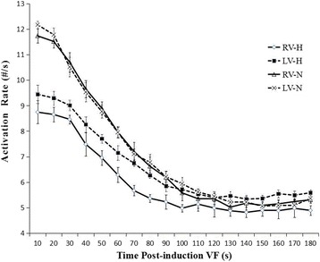
In the control group, the LV was activated faster than the RV in the first 20 s, after which there was no detectable difference in the AR between them. When analyzing the distribution of the AR within the bi-ventricles at 3 min of LDVF, the posterior LV was activated fastest, while the anterior was slowest. In the HF group, a detectable AR gradient existed between the two ventricles within 3 min of VF, with the LV activating more quickly than the RV. When analyzing the distribution of the AR within the bi-ventricles at 3 min of LDVF, the septum of the LV was activated fastest, while the anterior was activated slowest.
A global bi-ventricular endocardial AR gradient existed within the first 20 s of VF but disappeared in the LDVF in healthy hearts. However, the AR gradient was always observed in both SDVF and LDVF in HF hearts. The findings of this study suggest that LDVF in HF hearts can be maintained differently from normal hearts, which accordingly should lead to the development of different management strategies for LDVF resuscitation.
本研究的目的是检测正常和心力衰竭(HF)犬心脏在短程室颤(SDVF,<1分钟)和长程室颤(LDVF,>1分钟)期间左、右心室(LV & RV)激活率(AR)分布的差异。
对6只健康犬(对照组)和6只通过右心室起搏诱导充血性HF的犬(HF组)进行电诱导室颤(VF)。将两根64电极篮状导管分别置于左、右心室内,用于全心内膜电标测。通过快速傅里叶变换分析每个电极的VF的AR。
在对照组中,左心室在前20秒比右心室激活更快,此后两者之间的AR无明显差异。在分析LDVF 3分钟时双心室内AR的分布时,左心室后壁激活最快,而前壁最慢。在HF组中,VF 3分钟内两心室之间存在可检测到的AR梯度,左心室比右心室激活更快。在分析LDVF 3分钟时双心室内AR的分布时,左心室间隔激活最快,而前壁激活最慢。
VF最初20秒内存在全心内膜双心室AR梯度,但在健康心脏的LDVF中消失。然而,在HF心脏的SDVF和LDVF中均始终观察到AR梯度。本研究结果表明,HF心脏的LDVF维持方式与正常心脏不同,这相应地应导致针对LDVF复苏制定不同的管理策略。