Barbosa Flavia L, Xiao Yangyan, Bian Fang, Coursey Terry G, Ko Byung Yi, Clevers Hans, de Paiva Cintia S, Pflugfelder Stephen C
Department of Ophthalmology, Baylor College of Medicine, Houston, TX 77030, USA.
Department of Ophthalmology, the Second Xiangya Hospital, Central South University, Changsha 410011, China.
Int J Mol Sci. 2017 May 5;18(5):978. doi: 10.3390/ijms18050978.
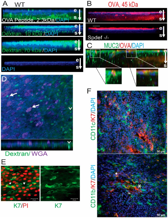
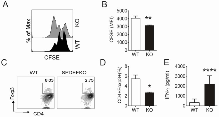
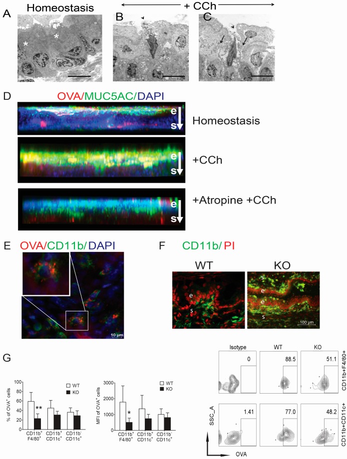
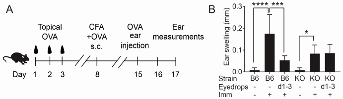
Conjunctival goblet cell (GC) loss in dry eye is associated with ocular surface inflammation. This study investigated if conjunctival GCs contribute to ocular surface immune tolerance. Antigens applied to the ocular surface, imaged by confocal microscopy, passed into the conjunctival stroma through goblet cell associated passages (GAPs) in wild type C57BL/6 (WT), while ovalbumin (OVA) was retained in the epithelium of SAM pointed domain containing ETS transcription factor () knockout mice (/) that lack GCs and are a novel model of dry eye. Stimulated GC degranulation increased antigen binding to GC mucins. Induction of tolerance to topically applied OVA measured by cutaneous delayed type hypersensitivity (DTH) was observed in WT, but not /. OTII CD4⁺ T cells primed by dendritic cells (DCs) from the conjunctival draining lymph nodes of / had greater IFN-γ production and lower Foxp3 positivity than those primed by WT DCs. These findings indicate that conjunctival GCs contribute to ocular surface immune tolerance by modulating antigen distribution and antigen specific immune response. GC loss may contribute to the abrogation of ocular surface immune tolerance that is observed in dry eye.
干眼患者结膜杯状细胞(GC)的丢失与眼表炎症有关。本研究调查结膜杯状细胞是否有助于眼表免疫耐受。通过共聚焦显微镜成像观察到,应用于眼表的抗原在野生型C57BL/6(WT)小鼠中通过杯状细胞相关通道(GAPs)进入结膜基质,而在缺乏杯状细胞且是一种新型干眼模型的含ETS转录因子()敲除小鼠(/)中,卵清蛋白(OVA)则保留在上皮中。刺激的杯状细胞脱颗粒增加了抗原与杯状细胞黏蛋白的结合。通过皮肤迟发型超敏反应(DTH)检测,在WT小鼠中观察到对局部应用OVA的耐受性诱导,但在/小鼠中未观察到。与由WT树突状细胞(DCs)致敏的OTII CD4⁺ T细胞相比,由/小鼠结膜引流淋巴结的DCs致敏的OTII CD4⁺ T细胞产生更多的IFN-γ且Foxp3阳性率更低。这些发现表明结膜杯状细胞通过调节抗原分布和抗原特异性免疫反应来促进眼表免疫耐受。杯状细胞丢失可能导致干眼患者眼表免疫耐受的丧失。