• 文献检索
  • 文档翻译
  • 深度研究
  • 学术资讯
  • Suppr Zotero 插件Zotero 插件
  • 邀请有礼
  • 套餐&价格
  • 历史记录
应用&插件
Suppr Zotero 插件Zotero 插件浏览器插件Mac 客户端Windows 客户端微信小程序
定价
高级版会员购买积分包购买API积分包
服务
文献检索文档翻译深度研究API 文档MCP 服务
关于我们
关于 Suppr公司介绍联系我们用户协议隐私条款
关注我们

Suppr 超能文献

核心技术专利:CN118964589B侵权必究
粤ICP备2023148730 号-1Suppr @ 2026

文献检索

告别复杂PubMed语法,用中文像聊天一样搜索,搜遍4000万医学文献。AI智能推荐,让科研检索更轻松。

立即免费搜索

文件翻译

保留排版,准确专业,支持PDF/Word/PPT等文件格式,支持 12+语言互译。

免费翻译文档

深度研究

AI帮你快速写综述,25分钟生成高质量综述,智能提取关键信息,辅助科研写作。

立即免费体验

通过CT滴注胆管造影术对副肝管进行术前评估。

Preoperative evaluation of accessory hepatic ducts by drip infusion cholangiography with CT.

作者信息

Ishii Hiromichi, Noguchi Akinori, Fukami Tomoyuki, Sugimoto Riho, Tada Hiroyuki, Takeshita Hiroki, Umehara Seiji, Izumi Hiroyuki, Tani Naoki, Yamaguchi Masahide, Yamane Tetsuro

机构信息

Department of Surgery, Matsushita Memorial Hospital, 5-55 Sotojima-cho, Moriguchi city, Osaka, 570-8540, Japan.

出版信息

BMC Surg. 2017 May 8;17(1):52. doi: 10.1186/s12893-017-0251-9.

DOI:10.1186/s12893-017-0251-9
PMID:28482819
原文链接:https://pmc.ncbi.nlm.nih.gov/articles/PMC5422935/
Abstract

BACKGROUND

This retrospective study aimed to investigate the incidence of each type of accessory hepatic duct by drip infusion cholangiography with CT (DIC-CT).

METHODS

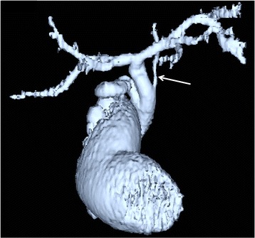
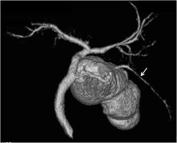
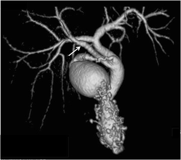
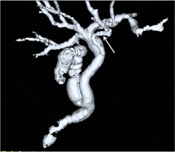
Five hundred sixty nine patients who underwent preoperative DIC-CT and laparoscopic cholecystectomy were reviewed. Accessory hepatic ducts were classified as follows: type I (accessory hepatic ducts that merged with the common hepatic duct between the confluence of the right and left hepatic ducts and the cystic duct confluence), type II (those that merged with the common hepatic duct at the same site as the cystic duct), type III (those that merged with the common bile duct distal to the cystic duct confluence), type IV (the cystic duct merged with the accessory hepatic duct), and type V (accessory hepatic ducts that merged with the common hepatic or bile duct on the left side).

RESULTS

Accessory hepatic ducts were observed in 50 patients. Type I, II, III, IV, and V accessory hepatic ducts were detected in 32, 3, 1, 11, and 3 patients, respectively. Based on their drainage areas, the accessory hepatic ducts were also classified as follows: a posterior branch in 22 patients, an anterior branch in 9 patients, a combination of posterior and anterior branches in 16 patients, a left-sided branch in 2 patients, and a caudate branch in 1 patient. None of the patients with accessory hepatic ducts suffered bile duct injuries.

CONCLUSION

There are a number of variants of the accessory hepatic duct. DIC-CT is useful to detect the accessory hepatic duct.

摘要

背景

本回顾性研究旨在通过CT滴注胆管造影术(DIC-CT)调查各型副肝管的发生率。

方法

回顾性分析569例行术前DIC-CT及腹腔镜胆囊切除术的患者。副肝管分类如下:I型(副肝管在左右肝管汇合处与胆囊管汇合处之间汇入肝总管),II型(副肝管在胆囊管汇入肝总管的同一部位汇入肝总管),III型(副肝管在胆囊管汇合处以远汇入胆总管),IV型(胆囊管汇入副肝管),V型(副肝管在左侧汇入肝总管或胆总管)。

结果

50例患者观察到副肝管。分别在32、3、1、11和3例患者中检测到I、II、III、IV和V型副肝管。根据其引流区域,副肝管也分类如下:22例为后支,9例为前支,16例为后支与前支联合,2例为左侧支,1例为尾状叶支。所有有副肝管的患者均未发生胆管损伤。

结论

副肝管有多种变异类型。DIC-CT有助于检测副肝管。

https://cdn.ncbi.nlm.nih.gov/pmc/blobs/ac9d/5422935/1e700e0912d7/12893_2017_251_Fig7_HTML.jpg
https://cdn.ncbi.nlm.nih.gov/pmc/blobs/ac9d/5422935/cf44632666bf/12893_2017_251_Fig1_HTML.jpg
https://cdn.ncbi.nlm.nih.gov/pmc/blobs/ac9d/5422935/f90b3c660d4b/12893_2017_251_Fig2_HTML.jpg
https://cdn.ncbi.nlm.nih.gov/pmc/blobs/ac9d/5422935/885e26e84e00/12893_2017_251_Fig3_HTML.jpg
https://cdn.ncbi.nlm.nih.gov/pmc/blobs/ac9d/5422935/8e4a3b803b1c/12893_2017_251_Fig4_HTML.jpg
https://cdn.ncbi.nlm.nih.gov/pmc/blobs/ac9d/5422935/1545ab124c69/12893_2017_251_Fig5_HTML.jpg
https://cdn.ncbi.nlm.nih.gov/pmc/blobs/ac9d/5422935/94177b767918/12893_2017_251_Fig6_HTML.jpg
https://cdn.ncbi.nlm.nih.gov/pmc/blobs/ac9d/5422935/1e700e0912d7/12893_2017_251_Fig7_HTML.jpg

相似文献

1
Preoperative evaluation of accessory hepatic ducts by drip infusion cholangiography with CT.通过CT滴注胆管造影术对副肝管进行术前评估。
BMC Surg. 2017 May 8;17(1):52. doi: 10.1186/s12893-017-0251-9.
2
Preoperative evaluation of the extrahepatic bile duct structure for laparoscopic cholecystectomy.腹腔镜胆囊切除术肝外胆管结构的术前评估
Surg Endosc. 2006 Jul;20(7):1119-23. doi: 10.1007/s00464-005-0689-1. Epub 2006 May 13.
3
Distributing pattern of the bile duct of the caudate lobe on computed tomography with drip infusion cholangiography and its surgical significance.尾状叶胆管在CT滴注胆管造影上的分布模式及其手术意义。
Hepatogastroenterology. 2004 Jan-Feb;51(55):29-32.
4
Noninvasive imaging of the biliary tree before or after laparoscopic cholecystectomy: use of three-dimensional spiral CT cholangiography.腹腔镜胆囊切除术前后胆管树的无创成像:三维螺旋CT胆管造影的应用
AJR Am J Roentgenol. 1994 Jun;162(6):1331-5. doi: 10.2214/ajr.162.6.8191994.
5
[Laparoscopic cholecystectomy--accessory bile ducts].[腹腔镜胆囊切除术——副胆管]
Acta Med Croatica. 2003;57(2):105-9.
6
Spiegel's lobe bile ducts often drain into the right hepatic duct or its branches: study using drip-infusion cholangiography-computed tomography in 179 consecutive patients.施皮格尔叶胆管常汇入右肝管或其分支:对179例连续患者进行滴注胆管造影-计算机断层扫描研究。
World J Surg. 2004 Oct;28(10):1001-6. doi: 10.1007/s00268-004-7483-4. Epub 2004 Sep 29.
7
Heterogeneity of subvesical ducts or the ducts of Luschka: a study using drip-infusion cholangiography-computed tomography in patients and cadaver specimens.膀胱下导管或卢施卡导管的异质性:一项在患者和尸体标本中使用滴注式胆管造影计算机断层扫描的研究。
World J Surg. 2005 Feb;29(2):217-23. doi: 10.1007/s00268-004-7652-5.
8
Efficacy of multi-slice computed tomography cholangiography before laparoscopic cholecystectomy.多层螺旋CT胆管造影术在腹腔镜胆囊切除术之前的疗效
ANZ J Surg. 2006 Aug;76(8):693-5. doi: 10.1111/j.1445-2197.2006.03833.x.
9
Evaluation of biliary anatomy in the caudate lobe using drip infusion cholangiography-computed tomography.应用滴注式胆管造影计算机断层扫描评估尾状叶胆管解剖结构。
Abdom Radiol (NY). 2019 Mar;44(3):886-893. doi: 10.1007/s00261-018-1825-4.
10
[Evaluation of anomalous bile ducts using helical CT cholangiography].[螺旋CT胆管造影对胆管异常的评估]
Nihon Igaku Hoshasen Gakkai Zasshi. 1998 Oct;58(12):692-9.

引用本文的文献

1
Cholelithiasis With a Rare Branched Accessory Hepatic Duct: A Case Report.伴有罕见分支副肝管的胆结石:一例报告
Cureus. 2025 Apr 16;17(4):e82409. doi: 10.7759/cureus.82409. eCollection 2025 Apr.
2
Laparoscopic cholecystectomy for a gallbladder with a short cystic duct draining to the accessory right anterior hepatic duct using indocyanine green fluorescence imaging: A case report.使用吲哚菁绿荧光成像技术对胆囊管短且引流至右肝前副肝管的胆囊进行腹腔镜胆囊切除术:一例报告。
Int J Surg Case Rep. 2024 Aug;121:110014. doi: 10.1016/j.ijscr.2024.110014. Epub 2024 Jul 6.
3
Novel per-oral cholangioscopy for treatment of occult accessory hepatic duct stones.

本文引用的文献

1
Impact of intraoperative cholangiography on postoperative morbidity and readmission: analysis of the NSQIP database.术中胆管造影对术后发病率和再入院率的影响:国家外科质量改进计划(NSQIP)数据库分析
Surg Endosc. 2016 Dec;30(12):5395-5403. doi: 10.1007/s00464-016-4896-8. Epub 2016 Apr 22.
2
Relationship between the risk of bile duct injury during laparoscopic cholecystectomy and the types of preoperative magnetic resonance cholangiopancreatiocography (MRCP).腹腔镜胆囊切除术期间胆管损伤风险与术前磁共振胰胆管造影(MRCP)类型之间的关系
Korean J Hepatobiliary Pancreat Surg. 2012 Feb;16(1):17-23. doi: 10.14701/kjhbps.2012.16.1.17. Epub 2012 Feb 29.
3
新型经口胆管镜治疗隐匿性副肝管结石
Gastroenterol Rep (Oxf). 2023 Aug 30;11:goad051. doi: 10.1093/gastro/goad051. eCollection 2023.
4
Gallbladder cancer concomitant with autosomal dominant polycystic kidney disease: A case report.胆囊癌合并常染色体显性多囊肾病:一例报告。
Clin Case Rep. 2022 Dec 15;10(12):e6734. doi: 10.1002/ccr3.6734. eCollection 2022 Dec.
5
Application of three-dimensional visualization technology in the anatomical variations of hilar bile ducts in Chinese population.三维可视化技术在中国人群肝门部胆管解剖变异中的应用
Front Surg. 2022 Aug 2;9:934183. doi: 10.3389/fsurg.2022.934183. eCollection 2022.
6
Effectiveness and Clinical Value of Laparoscopic Cholecystectomy and Cholangiography in the Diagnosis of Biliary Calculi.腹腔镜胆囊切除术及胆管造影在胆石症诊断中的有效性及临床价值
Front Surg. 2022 Apr 27;9:880266. doi: 10.3389/fsurg.2022.880266. eCollection 2022.
7
Modified and dynamic intraoperativecholangiography during laparoscopic cholecystectomy in two patients with aberrant right posterior hepatic duct.两名右后肝管异常患者在腹腔镜胆囊切除术中的改良动态术中胆管造影
Radiol Case Rep. 2022 Apr 4;17(6):1843-1847. doi: 10.1016/j.radcr.2022.03.031. eCollection 2022 Jun.
8
Risk identification and technical modifications reduce the incidence of post-cholecystectomy bile leakage: analysis of 5675 laparoscopic cholecystectomies.风险识别和技术改进可降低胆囊切除术后胆漏的发生率:5675 例腹腔镜胆囊切除术分析。
Langenbecks Arch Surg. 2022 Feb;407(1):213-223. doi: 10.1007/s00423-021-02264-z. Epub 2021 Aug 26.
9
Topographical relationship between the accessory hepatic duct and the hepatic artery system.副肝管与肝动脉系统的解剖位置关系。
Anat Sci Int. 2021 Jan;96(1):112-118. doi: 10.1007/s12565-020-00568-6. Epub 2020 Sep 11.
10
Risk of Gallstone Formation in Aberrant Extrahepatic Biliary Tract Anatomy: A Review of Literature.肝外胆管解剖异常时胆结石形成的风险:文献综述
Cureus. 2020 Aug 25;12(8):e10009. doi: 10.7759/cureus.10009.
Utility of fluorescent cholangiography during laparoscopic cholecystectomy: A systematic review.
荧光胆管造影在腹腔镜胆囊切除术中的应用:一项系统评价。
World J Gastroenterol. 2015 Jul 7;21(25):7877-83. doi: 10.3748/wjg.v21.i25.7877.
4
True left-sided gallbladder with variations of bile duct and cholecystic vein.真性左侧胆囊伴胆管及胆囊静脉变异
World J Gastroenterol. 2015 Jun 7;21(21):6754-8. doi: 10.3748/wjg.v21.i21.6754.
5
Preoperative detection and handling of aberrant right posterior sectoral hepatic duct during laparoscopic cholecystectomy.腹腔镜胆囊切除术中异常右后肝段胆管的术前检测与处理
J Hepatobiliary Pancreat Sci. 2015 Jul;22(7):558-62. doi: 10.1002/jhbp.252. Epub 2015 Apr 16.
6
Is intraoperative cholangiography necessary during laparoscopic cholecystectomy for cholelithiasis?在腹腔镜胆囊切除术治疗胆结石过程中,术中胆管造影是否必要?
World J Gastroenterol. 2015 Feb 21;21(7):2147-51. doi: 10.3748/wjg.v21.i7.2147.
7
Complications After Laparoscopic Cholecystectomy: A Video Evaluation Study of Whether the Critical View of Safety was Reached.腹腔镜胆囊切除术后的并发症:关于是否达到安全关键视野的视频评估研究
World J Surg. 2015 Jul;39(7):1798-803. doi: 10.1007/s00268-015-2993-9.
8
Correlation between portal vein anatomy and bile duct variation in 407 living liver donors.407例活体肝供体门静脉解剖与胆管变异的相关性
Am J Transplant. 2015 Jan;15(1):155-60. doi: 10.1111/ajt.12965. Epub 2014 Dec 17.
9
Three-dimensional imaging identified the accessory bile duct in a patient with cholangiocarcinoma.三维成像在一名胆管癌患者中识别出了副胆管。
World J Gastroenterol. 2014 Aug 28;20(32):11451-5. doi: 10.3748/wjg.v20.i32.11451.
10
CT and MR cholangiography: advantages and pitfalls in perioperative evaluation of biliary tree.CT 和 MR 胆管成像:胆道手术评估的优缺点。
Br J Radiol. 2012 Jul;85(1015):887-96. doi: 10.1259/bjr/21209407. Epub 2012 Mar 14.