Capasso Angelo, Raiano Vera, Sontuoso Antonio, Olivero Daniela, Greci Valentina
Gregory VII Veterinary Hospital, Rome, Italy.
Analisi BiEsseA Laboratory, Milan, Italy.
JFMS Open Rep. 2015 Jun 12;1(1):2055116915585019. doi: 10.1177/2055116915585019. eCollection 2015 Jan-Jun.
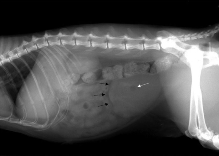
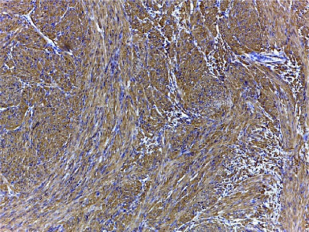
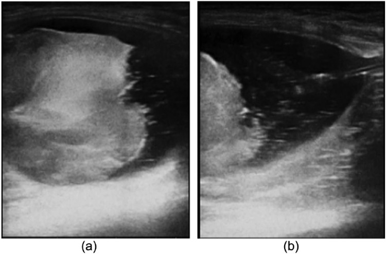
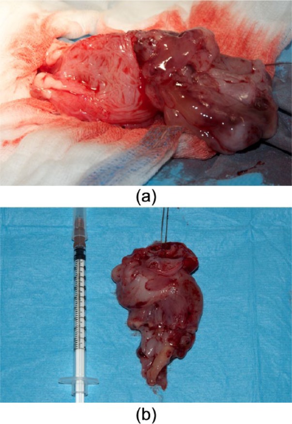
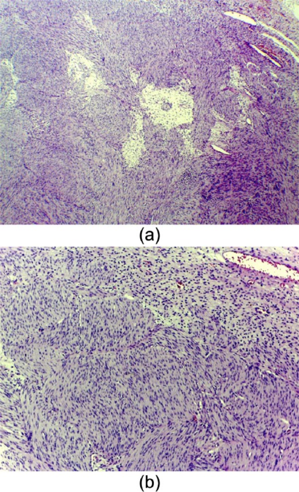
A 5-year-old female spayed domestic shorthair cat was presented with haematuria, pollakiuria and stranguria of 2 months' duration, and a firm non-painful mass in the urinary bladder was palpated. Abdominal radiographs showed thickening and irregular cranial margins of the urinary bladder wall. Abdominal ultrasound showed a vascularised mass of mixed echogenicity almost entirely occupying the urinary bladder lumen. During explorative laparotomy, the mass appeared pedunculated and was totally excised. Histopathology was characterised by infiltration of the mucosal, submucosal and muscular layers by proliferated atypical mesenchymal cells; immunochemistry confirmed the diagnosis of fibrosarcoma. The cat was discharged with normal urination 5 days after surgery. The owner declined any imaging follow-up but reported the cat to be free of any clinical signs at 16 months after surgery.
To the best of our knowledge, this is the first case of primary fibrosarcoma of the urinary bladder in the cat. Fibrosarcoma should be included in the differential diagnosis of urinary bladder neoplasia.
一只5岁已绝育的雌性家养短毛猫,出现持续2个月的血尿、尿频和排尿困难,触诊膀胱有一个坚硬、无压痛的肿块。腹部X线片显示膀胱壁增厚且颅侧边缘不规则。腹部超声显示一个混合回声的血管化肿块,几乎完全占据膀胱腔。在剖腹探查术中,肿块呈有蒂状,被完全切除。组织病理学特征为增生的非典型间充质细胞浸润黏膜、黏膜下层和肌层;免疫组化确诊为纤维肉瘤。术后5天猫排尿正常出院。主人拒绝任何影像学随访,但报告称术后16个月猫无任何临床症状。
据我们所知,这是猫原发性膀胱纤维肉瘤的首例病例。纤维肉瘤应纳入膀胱肿瘤的鉴别诊断。