Rasool R U, Nayak D, Chakraborty S, Faheem M M, Rah B, Mahajan P, Gopinath V, Katoch A, Iqra Z, Yousuf S K, Mukherjee D, Kumar L D, Nargotra A, Goswami A
Academy of Scientific and Innovative Research (AcSIR), CSIR-Indian Institute of Integrative Medicine, Jammu, India.
Cancer Pharmacology Division, CSIR-Indian Institute of Integrative Medicine, Jammu, India.
Oncogenesis. 2017 May 22;6(5):e341. doi: 10.1038/oncsis.2017.41.
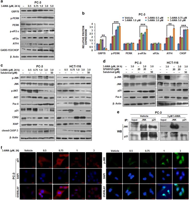
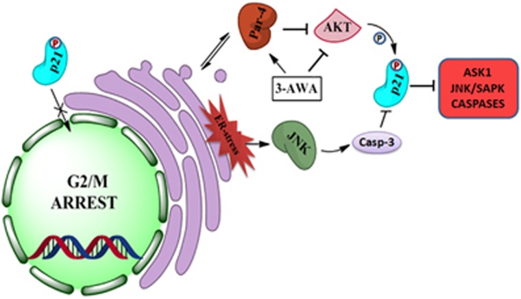
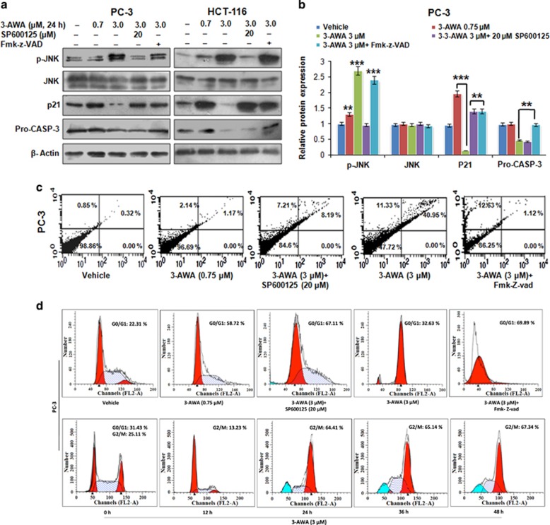
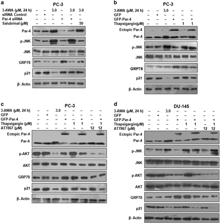
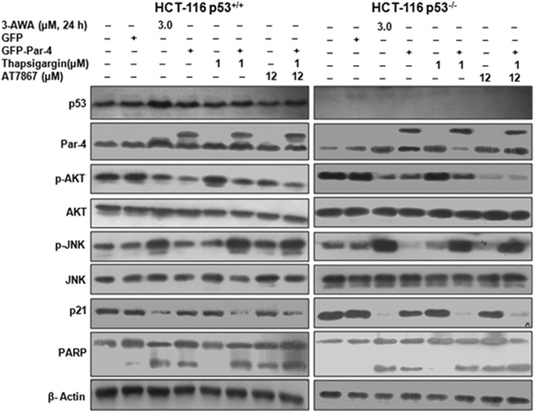
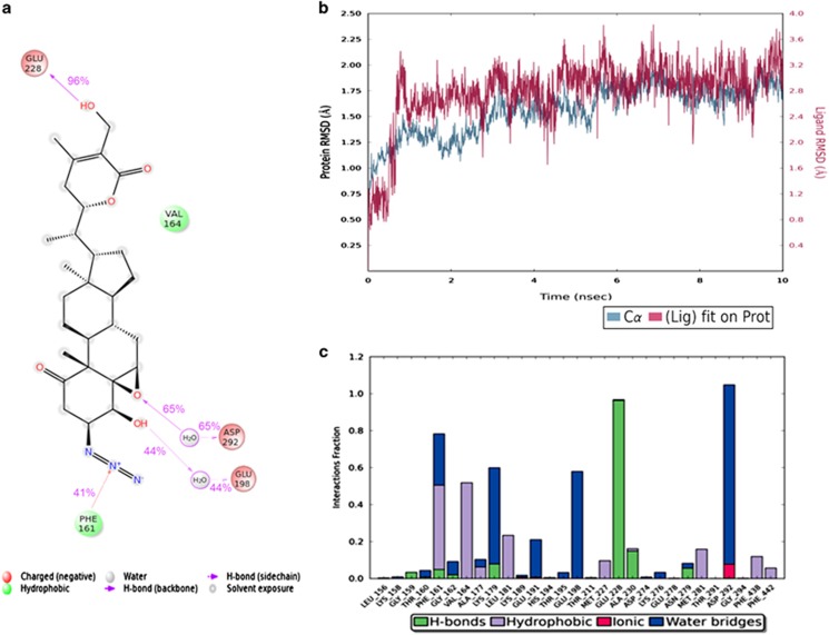
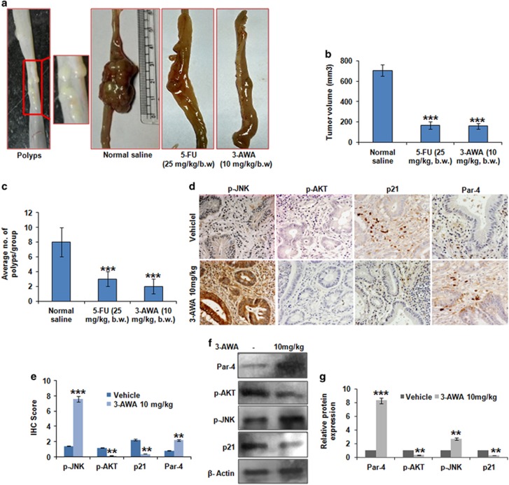
The double-edged role of p21 to command survival and apoptosis is emerging. The current investigation highlights ER stress-mediated JNK activation that plausibly triggers cell death by attenuating endogenous p21 level. Here, we demonstrated that ER stress activator 3-AWA diminishes the p21 levels in cancer cells by averting the senescent phenotype to commence G2/M arrest. In essence, the deceleration in p21 level occurs through ER stress/JNK/Caspase-3 axis via activation/induction of proapoptotic Par-4 and inhibition of AKT. The molecular dynamics studies identified important interactions, which may be responsible for the AKT inhibition and efficacy of 3-AWA towards AKT binding pocket. Interestingly, the p21 deceleration was rescued by incubating the cells with 3-AWA in the presence of an ER stress inhibitor, Salubrinal. Furthermore, we demonstrated that p21 expression decreases solitarily in Par-4 MEFs; albeit, ER stress-induced JNK activation was observed in both Par-4 and Par-4 MEFs. Par-4 knockdown or overexpression studies established that ectopic Par-4 along with ER stress are not sufficient to downregulate p21 in PC-3 cells but are adequate for DU-145 cells and that the ER stress inflicted activation of JNK, inhibition of AKT and Par-4 induction are all crucial to p21 downmodulation by 3-AWA. By using isogenic cell lines, such as HCT-116 p53 and HCT-116 p53, we found that deceleration in p21 expression due to ER stress is p53 independent. Moreover, in orthotopic carcinogen-induced rat colorectal carcinoma model, we found that 3-AWA inhibits colorectal tumor growth and formation of colorectal polyps at a tolerable dose, similar to the first-line drug for colorectal cancer-5-fluorouracil.
p21在调控细胞存活与凋亡方面的双重作用正逐渐显现。当前研究突出了内质网应激介导的JNK激活,这可能通过降低内源性p21水平来触发细胞死亡。在此,我们证明内质网应激激活剂3 - AWA通过避免衰老表型以启动G2/M期阻滞,从而降低癌细胞中的p21水平。本质上,p21水平的降低是通过内质网应激/JNK/半胱天冬酶 - 3轴,经由促凋亡蛋白Par - 4的激活/诱导以及AKT的抑制实现的。分子动力学研究确定了重要相互作用,这可能是AKT受到抑制以及3 - AWA对AKT结合口袋具有效力的原因。有趣的是,在存在内质网应激抑制剂Salubrinal的情况下,用3 - AWA处理细胞可挽救p21水平的降低。此外,我们证明p21表达仅在Par - 4基因敲除的小鼠胚胎成纤维细胞(MEFs)中降低;尽管如此,在Par - 4基因敲除的MEFs和野生型MEFs中均观察到内质网应激诱导的JNK激活。Par - 4基因敲低或过表达研究表明,异位表达的Par - 4与内质网应激一起并不足以下调PC - 3细胞中的p21,但对DU - 1细胞是足够的,并且内质网应激导致的JNK激活、AKT抑制和Par - 4诱导对于3 - AWA下调p21均至关重要。通过使用同基因细胞系,如HCT - 116 p53 +/+和HCT - 116 p53 -/-,我们发现内质网应激导致的p21表达降低与p53无关。此外,在原位致癌物诱导的大鼠结直肠癌模型中,我们发现3 - AWA在可耐受剂量下抑制结直肠癌肿瘤生长和结直肠息肉形成,类似于结直肠癌一线药物5 - 氟尿嘧啶。