• 文献检索
  • 文档翻译
  • 深度研究
  • 学术资讯
  • Suppr Zotero 插件Zotero 插件
  • 邀请有礼
  • 套餐&价格
  • 历史记录
应用&插件
Suppr Zotero 插件Zotero 插件浏览器插件Mac 客户端Windows 客户端微信小程序
定价
高级版会员购买积分包购买API积分包
服务
文献检索文档翻译深度研究API 文档MCP 服务
关于我们
关于 Suppr公司介绍联系我们用户协议隐私条款
关注我们

Suppr 超能文献

核心技术专利:CN118964589B侵权必究
粤ICP备2023148730 号-1Suppr @ 2026

文献检索

告别复杂PubMed语法,用中文像聊天一样搜索,搜遍4000万医学文献。AI智能推荐,让科研检索更轻松。

立即免费搜索

文件翻译

保留排版,准确专业,支持PDF/Word/PPT等文件格式,支持 12+语言互译。

免费翻译文档

深度研究

AI帮你快速写综述,25分钟生成高质量综述,智能提取关键信息,辅助科研写作。

立即免费体验

基于K均值聚类与改进分水岭算法的新组合从显微图像中分割白细胞

Segmentation of White Blood Cells From Microscopic Images Using a Novel Combination of K-Means Clustering and Modified Watershed Algorithm.

作者信息

Ghane Narjes, Vard Alireza, Talebi Ardeshir, Nematollahy Pardis

机构信息

Department of Bioelectrics and Biomedical Engineering, School of Advanced Technologies in Medicine and Student Research Center, Isfahan, Iran.

Department of Bioelectrics and Biomedical Engineering, School of Advanced Technologies in Medicine and Medical Image and Signal Processing Research Center, Isfahan, Iran.

出版信息

J Med Signals Sens. 2017 Apr-Jun;7(2):92-101.

PMID:28553582
原文链接:https://pmc.ncbi.nlm.nih.gov/articles/PMC5437768/
Abstract

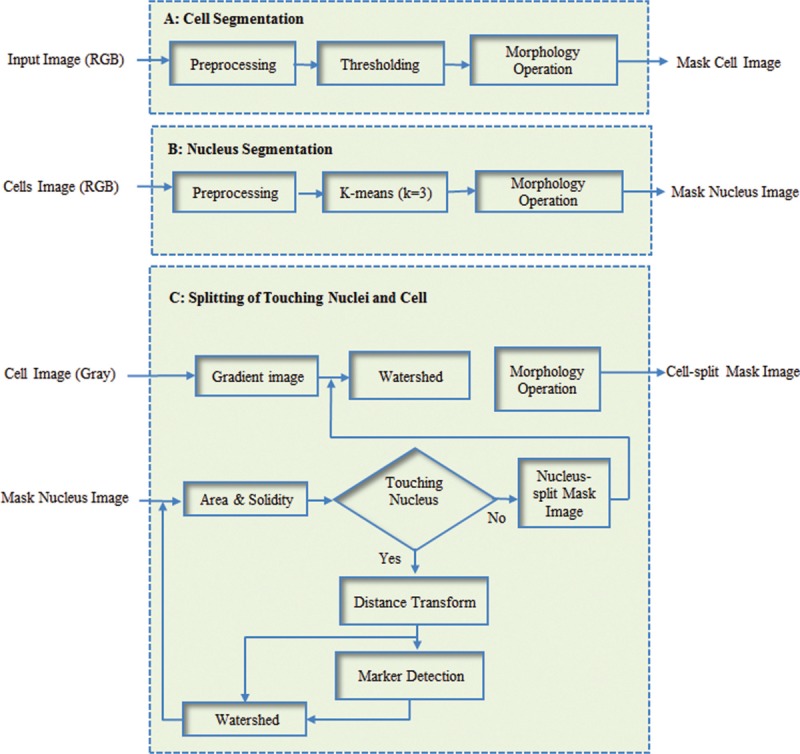
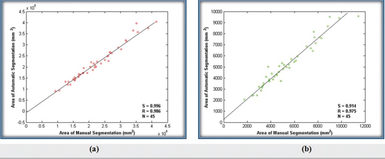
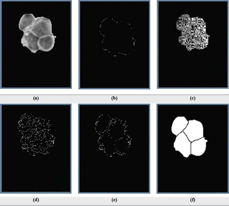
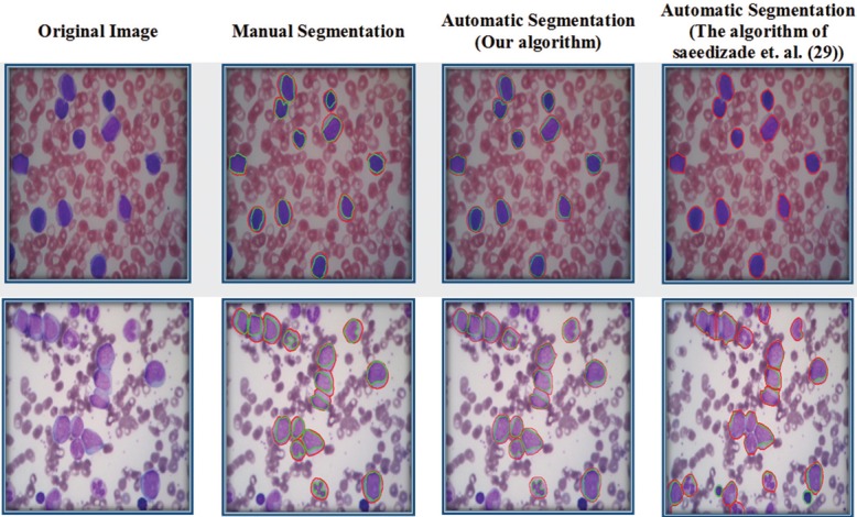
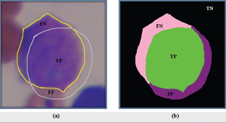
Recognition of white blood cells (WBCs) is the first step to diagnose some particular diseases such as acquired immune deficiency syndrome, leukemia, and other blood-related diseases that are usually done by pathologists using an optical microscope. This process is time-consuming, extremely tedious, and expensive and needs experienced experts in this field. Thus, a computer-aided diagnosis system that assists pathologists in the diagnostic process can be so effective. Segmentation of WBCs is usually a first step in developing a computer-aided diagnosis system. The main purpose of this paper is to segment WBCs from microscopic images. For this purpose, we present a novel combination of thresholding, k-means clustering, and modified watershed algorithms in three stages including (1) segmentation of WBCs from a microscopic image, (2) extraction of nuclei from cell's image, and (3) separation of overlapping cells and nuclei. The evaluation results of the proposed method show that similarity measures, precision, and sensitivity respectively were 92.07, 96.07, and 94.30% for nucleus segmentation and 92.93, 97.41, and 93.78% for cell segmentation. In addition, statistical analysis presents high similarity between manual segmentation and the results obtained by the proposed method.

摘要

识别白细胞是诊断某些特定疾病(如获得性免疫缺陷综合征、白血病和其他血液相关疾病)的第一步,这一过程通常由病理学家使用光学显微镜来完成。这个过程既耗时、极其繁琐又昂贵,并且需要该领域经验丰富的专家。因此,一个在诊断过程中协助病理学家的计算机辅助诊断系统会非常有效。白细胞分割通常是开发计算机辅助诊断系统的第一步。本文的主要目的是从微观图像中分割白细胞。为此,我们在三个阶段提出了一种新颖的阈值处理、k均值聚类和改进的分水岭算法的组合,包括(1)从微观图像中分割白细胞,(2)从细胞图像中提取细胞核,以及(3)分离重叠的细胞和细胞核。所提方法的评估结果表明,细胞核分割的相似性度量、精度和灵敏度分别为92.07%、96.07%和94.30%,细胞分割的相似性度量、精度和灵敏度分别为92.93%、97.41%和93.78%。此外,统计分析表明手动分割与所提方法获得的结果之间具有高度相似性。

https://cdn.ncbi.nlm.nih.gov/pmc/blobs/ce20/5437768/8f7c19ec5f9e/JMSS-7-92-g010.jpg
https://cdn.ncbi.nlm.nih.gov/pmc/blobs/ce20/5437768/4229df7863cd/JMSS-7-92-g001.jpg
https://cdn.ncbi.nlm.nih.gov/pmc/blobs/ce20/5437768/0102cc182e5c/JMSS-7-92-g002.jpg
https://cdn.ncbi.nlm.nih.gov/pmc/blobs/ce20/5437768/731418129b56/JMSS-7-92-g003.jpg
https://cdn.ncbi.nlm.nih.gov/pmc/blobs/ce20/5437768/369a04ab8166/JMSS-7-92-g005.jpg
https://cdn.ncbi.nlm.nih.gov/pmc/blobs/ce20/5437768/8d775d1098e1/JMSS-7-92-g006.jpg
https://cdn.ncbi.nlm.nih.gov/pmc/blobs/ce20/5437768/3a34fc15b0f6/JMSS-7-92-g007.jpg
https://cdn.ncbi.nlm.nih.gov/pmc/blobs/ce20/5437768/9a34a00c55cf/JMSS-7-92-g009.jpg
https://cdn.ncbi.nlm.nih.gov/pmc/blobs/ce20/5437768/8f7c19ec5f9e/JMSS-7-92-g010.jpg

相似文献

1
Segmentation of White Blood Cells From Microscopic Images Using a Novel Combination of K-Means Clustering and Modified Watershed Algorithm.基于K均值聚类与改进分水岭算法的新组合从显微图像中分割白细胞
J Med Signals Sens. 2017 Apr-Jun;7(2):92-101.
2
WBC-based segmentation and classification on microscopic images: a minor improvement.基于白细胞的显微镜图像分割与分类:略有改进。
F1000Res. 2021 Nov 17;10:1168. doi: 10.12688/f1000research.73315.1. eCollection 2021.
3
Nucleus and cytoplasm segmentation in microscopic images using K-means clustering and region growing.使用K均值聚类和区域生长法对微观图像中的细胞核与细胞质进行分割
Adv Biomed Res. 2015 Aug 31;4:174. doi: 10.4103/2277-9175.163998. eCollection 2015.
4
Segmentation of white blood cells and comparison of cell morphology by linear and naïve Bayes classifiers.白细胞的分割以及通过线性和朴素贝叶斯分类器对细胞形态的比较。
Biomed Eng Online. 2015 Jun 30;14:63. doi: 10.1186/s12938-015-0037-1.
5
A novel white blood cells segmentation algorithm based on adaptive neutrosophic similarity score.一种基于自适应中智相似性分数的新型白细胞分割算法。
Health Inf Sci Syst. 2017 Dec 18;6(1):1. doi: 10.1007/s13755-017-0038-5. eCollection 2018 Dec.
6
Fast unsupervised nuclear segmentation and classification scheme for automatic allred cancer scoring in immunohistochemical breast tissue images.快速无监督的核分割和分类方案,用于免疫组织化学乳腺组织图像中的自动 Allred 癌症评分。
Comput Methods Programs Biomed. 2018 Oct;165:37-51. doi: 10.1016/j.cmpb.2018.08.005. Epub 2018 Aug 10.
7
Automatic detection and quantification of WBCs and RBCs using iterative structured circle detection algorithm.利用迭代结构圆检测算法自动检测和量化白细胞和红细胞。
Comput Math Methods Med. 2014;2014:979302. doi: 10.1155/2014/979302. Epub 2014 Apr 3.
8
White blood cell segmentation by color-space-based k-means clustering.基于颜色空间的k均值聚类进行白细胞分割
Sensors (Basel). 2014 Sep 1;14(9):16128-47. doi: 10.3390/s140916128.
9
Segmentation of White Blood Cells through Nucleus Mark Watershed Operations and Mean Shift Clustering.通过细胞核标记分水岭算法和均值漂移聚类实现白细胞分割
Sensors (Basel). 2015 Sep 8;15(9):22561-86. doi: 10.3390/s150922561.
10
Nucleus segmentation of white blood cells in blood smear images by modeling the pixels' intensities as a set of three Gaussian distributions.通过将像素的强度建模为三个高斯分布的集合来对血涂片图像中的白细胞进行核分割。
Med Biol Eng Comput. 2024 Aug;62(8):2371-2388. doi: 10.1007/s11517-024-03065-4. Epub 2024 Apr 8.

引用本文的文献

1
Cell Nuclear Segmentation of B-ALL Images Based on MSFF-SegNeXt.基于MSFF-SegNeXt的B-ALL图像细胞核分割
J Multidiscip Healthc. 2024 Dec 2;17:5675-5693. doi: 10.2147/JMDH.S492655. eCollection 2024.
2
Threshold estimation based on local minima for nucleus and cytoplasm segmentation.基于局部极小值的细胞核和细胞质分割的阈值估计。
BMC Med Imaging. 2022 Apr 26;22(1):77. doi: 10.1186/s12880-022-00801-w.
3
WBC-based segmentation and classification on microscopic images: a minor improvement.基于白细胞的显微镜图像分割与分类:略有改进。

本文引用的文献

1
Robust Nucleus/Cell Detection and Segmentation in Digital Pathology and Microscopy Images: A Comprehensive Review.在数字病理学和显微镜图像中进行稳健的细胞核/细胞检测和分割:全面综述。
IEEE Rev Biomed Eng. 2016;9:234-63. doi: 10.1109/RBME.2016.2515127. Epub 2016 Jan 6.
2
Nucleus and cytoplasm segmentation in microscopic images using K-means clustering and region growing.使用K均值聚类和区域生长法对微观图像中的细胞核与细胞质进行分割
Adv Biomed Res. 2015 Aug 31;4:174. doi: 10.4103/2277-9175.163998. eCollection 2015.
3
Automatic recognition of myeloma cells in microscopic images using bottleneck algorithm, modified watershed and SVM classifier.
F1000Res. 2021 Nov 17;10:1168. doi: 10.12688/f1000research.73315.1. eCollection 2021.
4
New segmentation and feature extraction algorithm for classification of white blood cells in peripheral smear images.外周血涂片白细胞分类的新分割与特征提取算法。
Sci Rep. 2021 Sep 30;11(1):19428. doi: 10.1038/s41598-021-98599-0.
5
A Novel Method for Effective Cell Segmentation and Tracking in Phase Contrast Microscopic Images.一种用于相差显微镜图像中有效细胞分割和跟踪的新方法。
Sensors (Basel). 2021 May 18;21(10):3516. doi: 10.3390/s21103516.
6
Classification of chronic myeloid leukemia cell subtypes based on microscopic image analysis.基于显微图像分析的慢性髓性白血病细胞亚型分类
EXCLI J. 2019 Jun 14;18:382-404. doi: 10.17179/excli2019-1292. eCollection 2019.
7
Image Processing Approach for Detection of Leukocytes in Peripheral Blood Smears.图像处理方法在外周血涂片白细胞检测中的应用。
J Med Syst. 2019 Mar 22;43(5):114. doi: 10.1007/s10916-019-1219-3.
8
A novel white blood cells segmentation algorithm based on adaptive neutrosophic similarity score.一种基于自适应中智相似性分数的新型白细胞分割算法。
Health Inf Sci Syst. 2017 Dec 18;6(1):1. doi: 10.1007/s13755-017-0038-5. eCollection 2018 Dec.
使用瓶颈算法、改进的分水岭算法和支持向量机分类器自动识别显微图像中的骨髓瘤细胞。
J Microsc. 2016 Jan;261(1):46-56. doi: 10.1111/jmi.12314. Epub 2015 Oct 12.
4
Leucocyte classification for leukaemia detection using image processing techniques.利用图像处理技术进行白血病检测的白细胞分类
Artif Intell Med. 2014 Nov;62(3):179-91. doi: 10.1016/j.artmed.2014.09.002. Epub 2014 Sep 16.
5
Extraction of nucleolus candidate zone in white blood cells of peripheral blood smear images using curvelet transform.使用curvelet 变换提取外周血涂片白细胞核仁候选区。
Comput Math Methods Med. 2012;2012:574184. doi: 10.1155/2012/574184. Epub 2012 May 15.
6
An automated approach for segmentation of intravascular ultrasound images based on parametric active contour models.一种基于参数活动轮廓模型的血管内超声图像分割自动化方法。
Australas Phys Eng Sci Med. 2012 Jun;35(2):135-50. doi: 10.1007/s13246-012-0131-7. Epub 2012 Mar 14.
7
Automatic recognition of five types of white blood cells in peripheral blood.外周血中五种白细胞的自动识别。
Comput Med Imaging Graph. 2011 Jun;35(4):333-43. doi: 10.1016/j.compmedimag.2011.01.003.
8
Automated leukocyte recognition using fuzzy divergence.基于模糊散度的白细胞自动识别
Micron. 2010 Oct;41(7):840-6. doi: 10.1016/j.micron.2010.04.017. Epub 2010 May 8.
9
A framework for white blood cell segmentation in microscopic blood images using digital image processing.利用数字图像处理技术对显微镜下的血图像中的白细胞进行分割的框架。
Biol Proced Online. 2009 Jun 11;11:196-206. doi: 10.1007/s12575-009-9011-2.
10
Unsupervised segmentation based on robust estimation and color active contour models.基于稳健估计和彩色活动轮廓模型的无监督分割
IEEE Trans Inf Technol Biomed. 2005 Sep;9(3):475-86. doi: 10.1109/titb.2005.847515.