Chen Donglei, Blom Henning, Sanchez Sophie, Tafforeau Paul, Märss Tiiu, Ahlberg Per E
Department of Organismal Biology, Uppsala University, Norbyvägen 18A, 752 36, Uppsala, Sweden.
SciLifeLab, Uppsala University, Norbyvägen 18A, 752 36, Uppsala, Sweden.
R Soc Open Sci. 2017 May 17;4(5):161084. doi: 10.1098/rsos.161084. eCollection 2017 May.
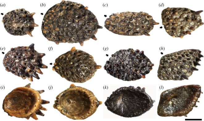
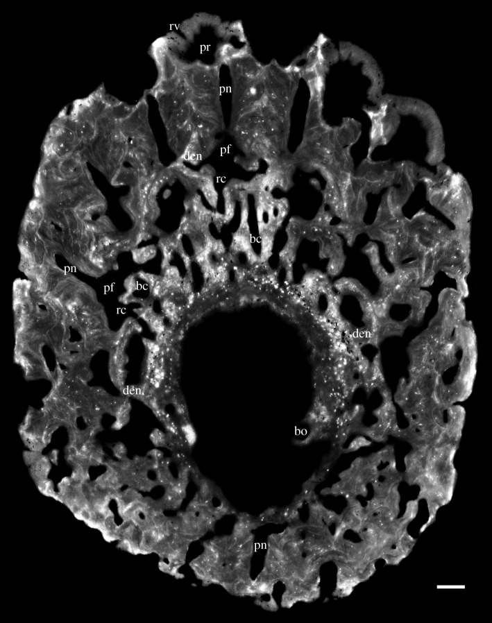
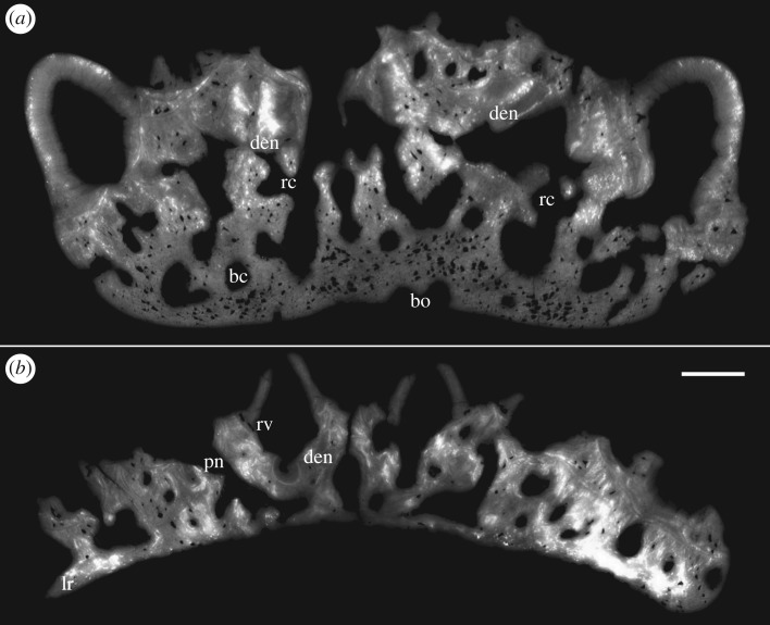
The numerous cushion-shaped tooth-bearing plates attributed to the stem group osteichthyan , which are argued here to represent an early form of the osteichthyan inner dental arcade, display a previously unknown and presumably primitive mode of tooth shedding by basal hard tissue resorption. They carry regularly spaced, recumbent, gently recurved teeth arranged in transverse tooth files that diverge towards the lingual margin of the cushion. Three-dimensional reconstruction from propagation phase-contrast synchrotron microtomography (PPC-SRµCT) reveals remnants of the first-generation teeth embedded in the basal plate, a feature never previously observed in any taxon. These teeth were shed by semi-basal resorption with the periphery of their bases retained as dentine rings. The rings are highly overlapped, which evidences tooth shedding prior to adding the next first-generation tooth at the growing edge of the plate. The first generation of teeth is thus diachronous. Successor teeth at the same sites underwent cyclical replacing and shedding through basal resorption, producing stacks of buried resorption surfaces separated by bone of attachment. The number and spatial arrangement of resorption surfaces elucidates that basal resorption of replacement teeth had taken place at the older tooth sites before the addition of the youngest first-generation teeth at the lingual margin. Thus, the replacement tooth buds cannot have been generated by a single permanent dental lamina at the lingual edge of the tooth cushion, but must have arisen either from successional dental laminae associated with the individual predecessor teeth, or directly from the dental epithelium of these teeth. The virtual histological dissection of these Late Silurian microfossils broadens our understanding of the development of the gnathostome dental systems and the acquisition of the osteichthyan-type of tooth replacement.
众多归因于硬骨鱼干群的垫状齿板,本文认为它们代表了硬骨鱼内牙弓的早期形式,通过基部硬组织吸收显示出一种前所未知且可能原始的牙齿脱落模式。它们带有规则间隔、平卧、微弯的牙齿,排列成横向齿列,向垫的舌缘发散。传播相衬同步辐射显微断层扫描(PPC-SRµCT)的三维重建揭示了嵌入基板的第一代牙齿的残余物,这是以前在任何分类群中从未观察到的特征。这些牙齿通过半基部吸收脱落,其基部周边保留为牙本质环。这些环高度重叠,这证明在板的生长边缘添加下一颗第一代牙齿之前牙齿已经脱落。因此,第一代牙齿是不同时的。同一部位的后继牙齿通过基部吸收进行周期性替换和脱落,产生由附着骨隔开的埋入吸收表面堆叠。吸收表面的数量和空间排列表明,替换牙齿的基部吸收在较老的牙齿部位发生,然后才在舌缘添加最年轻的第一代牙齿。因此,替换牙胚不可能由齿垫舌缘的单个恒牙板产生,而必定要么来自与单个前导牙齿相关的后继牙板,要么直接来自这些牙齿的牙上皮。这些志留纪晚期微化石的虚拟组织学剖析拓宽了我们对颌口动物牙齿系统发育以及硬骨鱼型牙齿替换获得的理解。