Takaoka Taiki, Shibamoto Yuta, Matsuo Masayuki, Sugie Chikao, Murai Taro, Ogawa Yasutaka, Miyakawa Akifumi, Manabe Yoshihiko, Kondo Takuhito, Nakajima Koichiro, Okazaki Dai, Tsuchiya Takahiro
Department of Radiology, Nagoya City University Graduate School of Medical Sciences, Nagoya, Japan.
Department of Radiology, Gifu University School of Medicine, Gifu, Japan.
Cancer Sci. 2017 Sep;108(9):1787-1792. doi: 10.1111/cas.13302. Epub 2017 Jul 21.
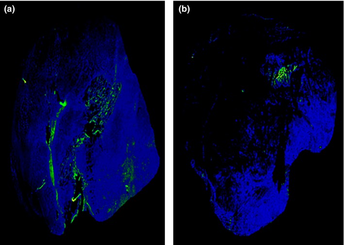
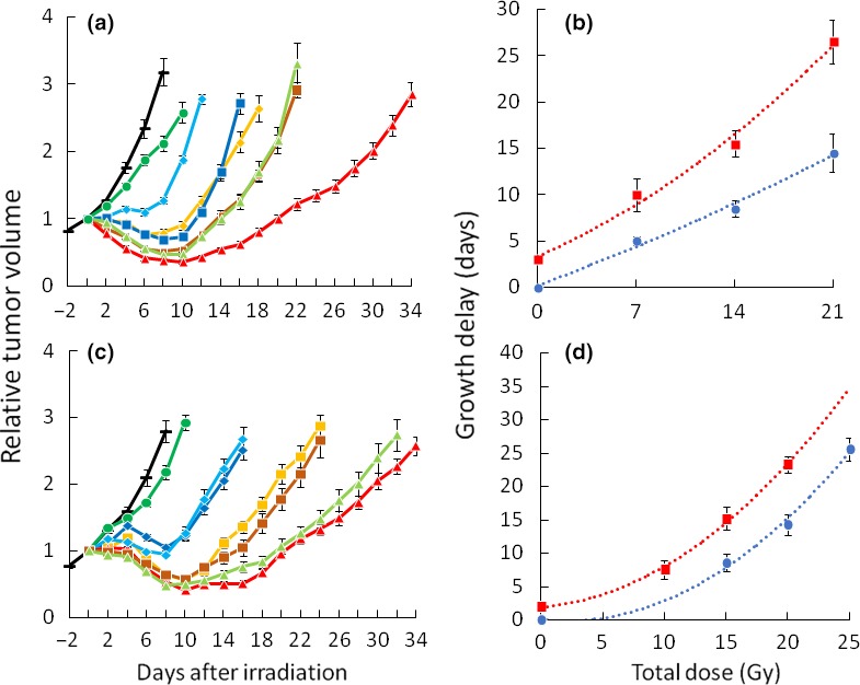
Despite insufficient laboratory data, radiotherapy after intratumoral injection of hydrogen peroxide (H O ) is increasingly being used clinically for radioresistant tumors. Especially, this treatment might become an alternative definitive treatment for early and advanced breast cancer in patients who refuse any type of surgery. The purpose of this study was to investigate the biological effects and appropriate combination methods of irradiation and H O in vivo. SCCVII tumor cells transplanted into the legs of C3H/HeN mice were used. Chronological changes of intratumoral distribution of oxygen bubbles after injection of H O were investigated using computed tomography. The effects of H O alone and in combination with single or five-fraction irradiation were investigated using a growth delay assay. The optimal timing of H O injection was investigated. Immunostaining of tumors was performed using the hypoxia marker pimonidazole. Oxygen bubbles decreased gradually and almost disappeared after 24 h. Administration of H O produced 2-3 days' tumor growth delay. Tumor regrowth was slowed further when H O was injected before irradiation. The group irradiated immediately after H O injection showed the longest tumor growth delay. Dose-modifying factors were 1.7-2.0 when combined with single irradiation and 1.3-1.5 with fractionated irradiation. Pimonidazole staining was weaker in tumors injected with H O . H O injection alone had modest antitumor effects. Greater tumor growth delays were demonstrated by combining irradiation and H O injection. The results of the present study could serve as a basis for evaluating results of various clinical studies on this treatment.
尽管实验室数据不足,但瘤内注射过氧化氢(H₂O₂)后的放射治疗在临床上越来越多地用于治疗放射抗拒性肿瘤。特别是,对于拒绝任何类型手术的早期和晚期乳腺癌患者,这种治疗可能成为一种替代的确定性治疗方法。本研究的目的是研究体内照射与H₂O₂的生物学效应及合适的联合方法。使用移植到C3H/HeN小鼠腿部的SCCVII肿瘤细胞。利用计算机断层扫描研究注射H₂O₂后瘤内氧气泡分布的时间变化。使用生长延迟试验研究单独使用H₂O₂以及与单次或五次分割照射联合使用的效果。研究H₂O₂注射的最佳时机。使用缺氧标记物匹莫硝唑对肿瘤进行免疫染色。氧气泡逐渐减少,24小时后几乎消失。给予H₂O₂可使肿瘤生长延迟2 - 3天。在照射前注射H₂O₂时,肿瘤再生长进一步减慢。在注射H₂O₂后立即照射的组显示出最长的肿瘤生长延迟。与单次照射联合时剂量修正因子为1.7 - 2.0,与分割照射联合时为1.3 - 1.5。在注射H₂O₂的肿瘤中匹莫硝唑染色较弱。单独注射H₂O₂具有适度的抗肿瘤作用。联合照射和注射H₂O₂显示出更大的肿瘤生长延迟。本研究结果可为评估该治疗的各种临床研究结果提供依据。