Eggermont Florieke, Derikx Loes C, Verdonschot Nico, Hannink Gerjon, Kaatee Robert S J P, Tanck Esther, van der Linden Yvette M
Orthopaedic Research Laboratory, Radboud Institute for Health Sciences, Radboud university medical center, Nijmegen, The Netherlands.
Laboratory of Biomechanical Engineering, University of Twente, Enschede, The Netherlands.
Adv Radiat Oncol. 2016 Nov 10;2(1):53-61. doi: 10.1016/j.adro.2016.11.001. eCollection 2017 Jan-Mar.
The aim of this study was to determine the effect of single fraction (SF) and multiple fraction (MF) radiation therapy (RT) on bone mineral density (BMD) in patients with cancer and bone metastases in the proximal femur. We studied this effect in the radiation field and within metastatic lesions, and differentiated between lytic, blastic, and mixed lesions.
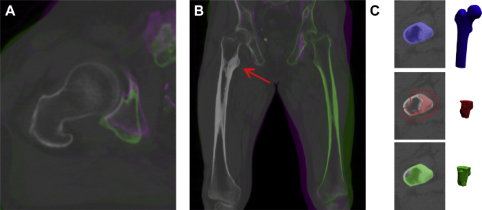
This prospective cohort study comprised 42 patients with painful bone metastases, including 47 irradiated femora with 52 metastatic lesions in the proximal femur. Patients received either 8 Gy SF or 20 to 24 Gy in 5 to 6 fractions (MF). Quantitative computed tomography scans were obtained before RT and 4 and 10 weeks after the initial scan. Patients who received MF additionally underwent quantitative computed tomography on the final day of their treatment. Automated image registration was performed. Mean BMD was determined at each time point for each proximal femur (region of interest [ROI]-PF) and in greater detail for a region of interest that contained the metastatic lesion (ROI-ML). Statistical analysis was performed using linear mixed models.
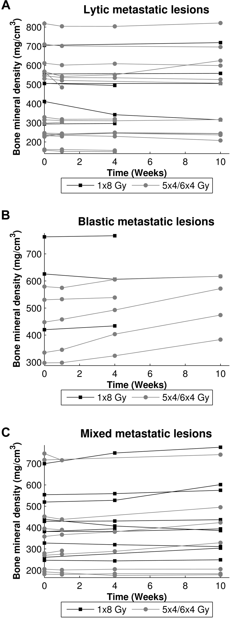
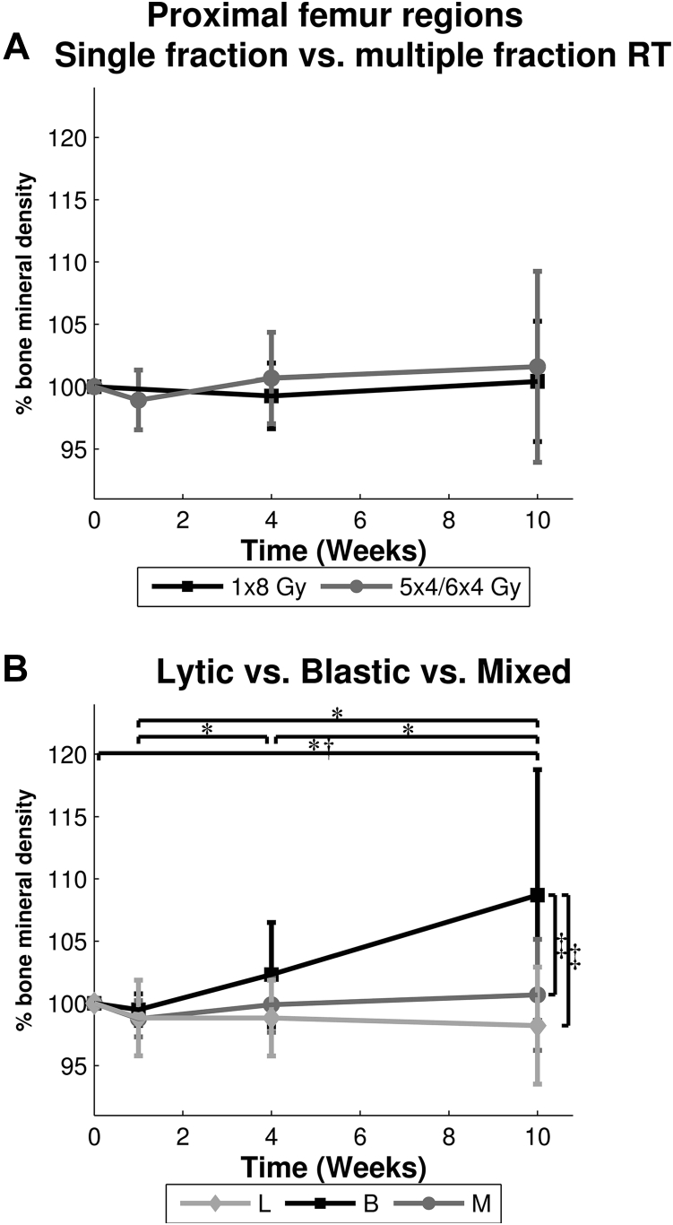
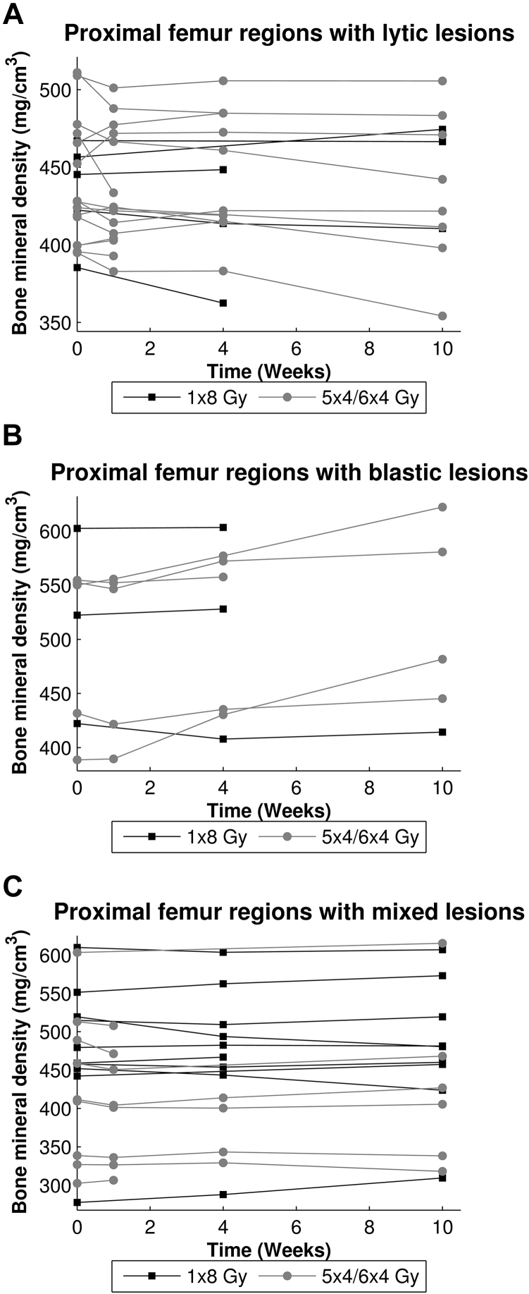
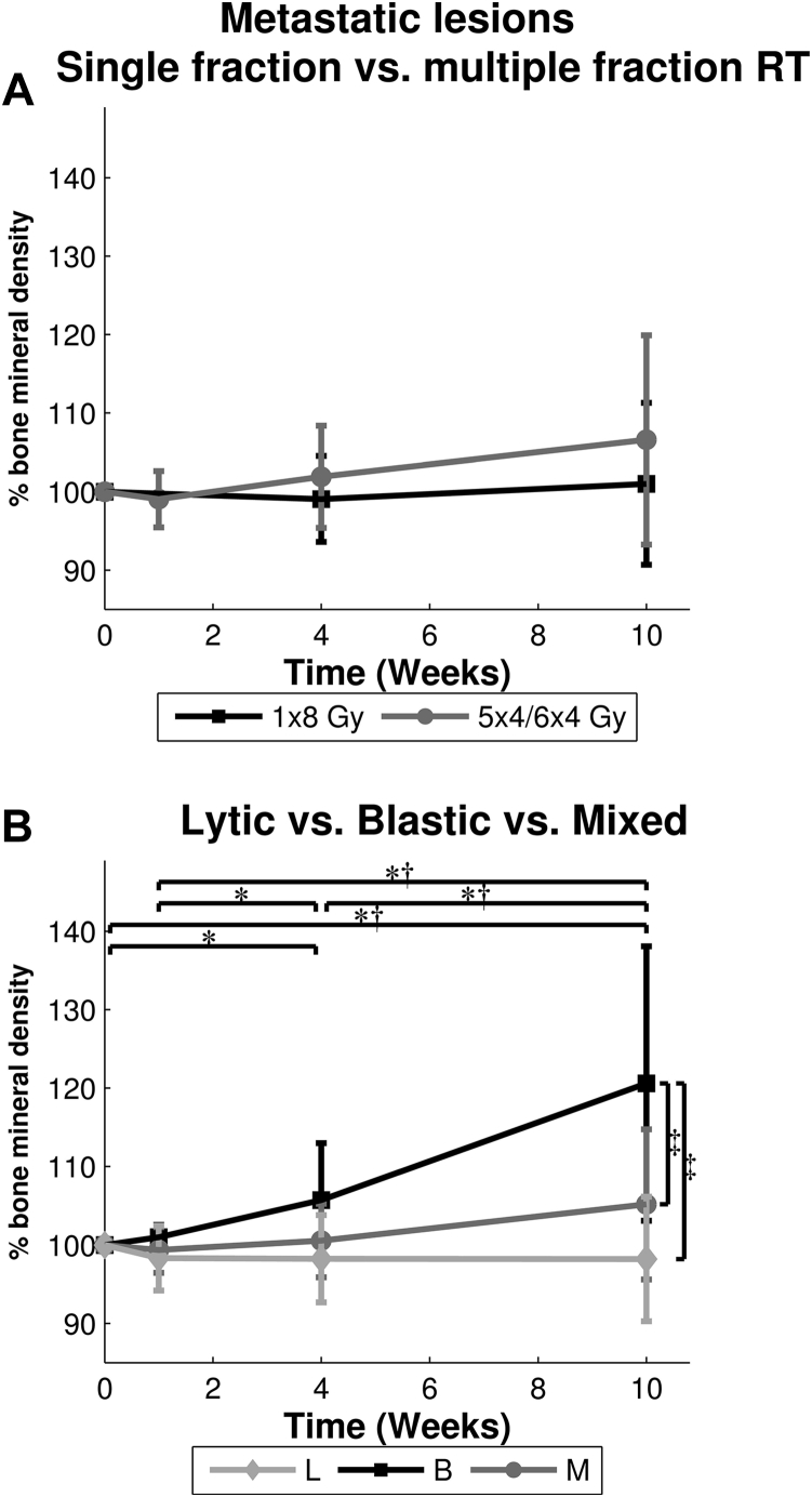
No significant differences in mean BMD were found between SF or MF RT over all time points in both ROI-PF and ROI-ML. Mean BMD did not change in ROI-PF with lytic and mixed lesions, but mean BMD in ROI-PF with blastic lesions increased to 109%. Comparably, when focused on ROI-ML, no differences in mean BMD were observed in lytic ROI-ML but mean BMD in mixed and blastic ROI-ML increased up to 105% and 121%, respectively.
Ten weeks after palliative radiation therapy in patients with femoral metastatic lesions, a limited increase in BMD was seen with no beneficial effect of MF over SF RT. BMD in lytic lesions was unchanged but slightly increased in mixed and blastic lesions.
本研究旨在确定单次分割(SF)和多次分割(MF)放射治疗(RT)对患有癌症且股骨近端发生骨转移的患者骨密度(BMD)的影响。我们在放射野内以及转移病灶内研究了这种影响,并区分了溶骨性、成骨性和混合性病灶。
这项前瞻性队列研究纳入了42例伴有疼痛性骨转移的患者,包括47例接受照射的股骨,其中52个转移病灶位于股骨近端。患者接受8 Gy的单次分割照射或20至24 Gy分5至6次分割(多次分割)照射。在放疗前以及首次扫描后的4周和10周进行定量计算机断层扫描。接受多次分割照射的患者在治疗的最后一天还额外进行了定量计算机断层扫描。进行了自动图像配准。在每个时间点确定每个股骨近端(感兴趣区域[ROI]-PF)的平均骨密度,并更详细地确定包含转移病灶的感兴趣区域(ROI-ML)的平均骨密度。使用线性混合模型进行统计分析。
在ROI-PF和ROI-ML的所有时间点上,单次分割或多次分割放疗之间的平均骨密度均未发现显著差异。溶骨性和混合性病灶的ROI-PF中的平均骨密度没有变化,但成骨性病灶的ROI-PF中的平均骨密度增加到了109%。同样,当关注ROI-ML时,溶骨性ROI-ML中的平均骨密度没有差异,但混合性和成骨性ROI-ML中的平均骨密度分别增加到了105%和121%。
在股骨转移病灶患者接受姑息性放疗10周后,骨密度有有限增加,多次分割放疗相对于单次分割放疗没有益处。溶骨性病灶中的骨密度没有变化,但混合性和成骨性病灶中的骨密度略有增加。