Halankar Jaydeep, Jhaveri Kartik, Metser Ur
Joint Department of Medical Imaging, University Health Network, Mount Sinai Hospital and Women's College Hospital, University of Toronto, Toronto, Ontario, Canada.
Indian J Radiol Imaging. 2017 Apr-Jun;27(2):167-176. doi: 10.4103/ijri.IJRI_226_16.
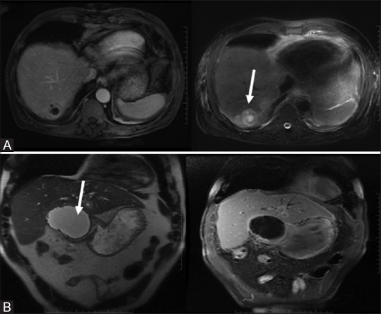
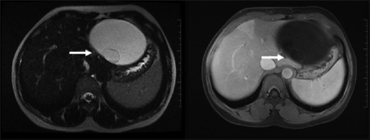
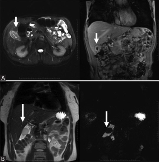
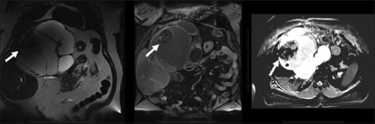
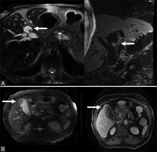
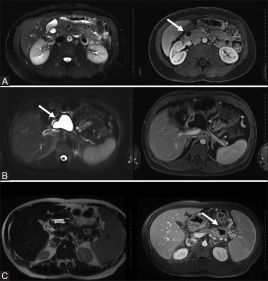
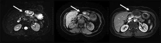
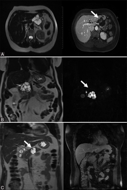
Although a common occurrence, cystic lesions of the pancreatico-biliary tree (PBT) may pose a diagnostic dilemma because they encompass a large number of neoplastic and benign processes with varied clinical symptoms. Knowledge of lesion classification and characterization are essential in making an accurate prospective diagnosis. This is necessary for identifying clinically significant cystic masses, which at times may require invasive intervention from indolent, nonneoplastic lesions, for which surveillance may suffice. Today, there is an arsenal of modalities for assessing the PBT, however, magnetic resonance imaging (MRI) remains at the forefront for characterizing cystic morphology and fluid content, internal septations, solid component, enhancement patterns, as well as assessing the surrounding normal structures. This pictorial review aims to review the spectrum of MRI features, which will aid in the differential diagnoses of cystic lesions of the PBT and mimickers, enabling the radiologist to reach a more confident diagnosis.
尽管胰胆管树(PBT)的囊性病变很常见,但可能会造成诊断困境,因为它们包含大量具有不同临床症状的肿瘤性和良性病变。了解病变的分类和特征对于做出准确的前瞻性诊断至关重要。这对于识别具有临床意义的囊性肿块是必要的,这些肿块有时可能需要对惰性的非肿瘤性病变进行侵入性干预,而对于后者,监测可能就足够了。如今,有大量用于评估PBT的检查方法,然而,磁共振成像(MRI)在表征囊性形态、液体成分、内部间隔、实性成分、强化模式以及评估周围正常结构方面仍处于领先地位。本图像综述旨在回顾MRI特征谱,这将有助于对PBT囊性病变及其模仿者进行鉴别诊断,使放射科医生能够做出更有把握的诊断。