Go Yoon Young, Kim Sung Eun, Cho Geum Joon, Chae Sung-Won, Song Jae-Jun
Department of Otorhinolaryngology-Head and Neck Surgery, Korea University College of Medicine, Seoul, Korea.
Department of Orthopedic Surgery and Rare Diseases Institute, Korea University College of Medicine, Seoul, Korea.
PLoS One. 2017 Aug 10;12(8):e0182716. doi: 10.1371/journal.pone.0182716. eCollection 2017.
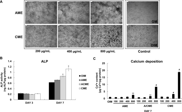
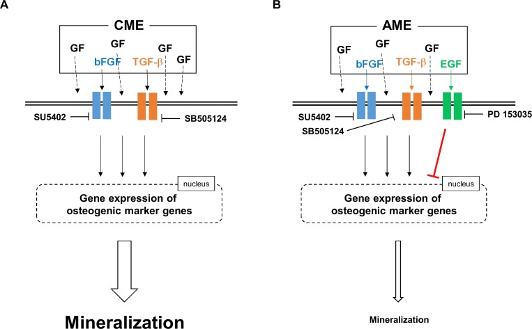
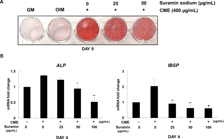
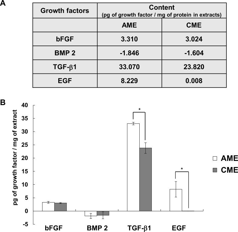
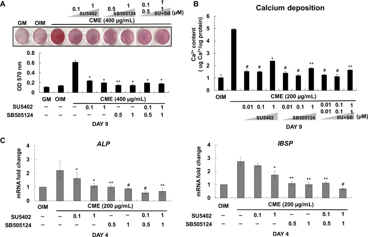
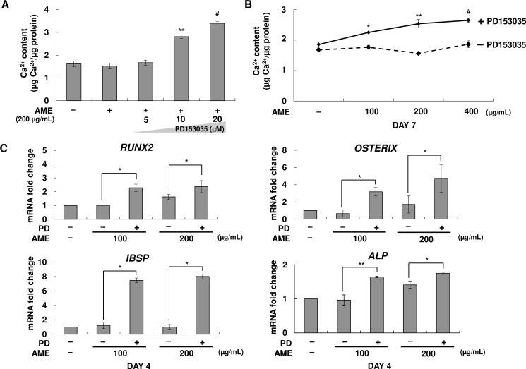
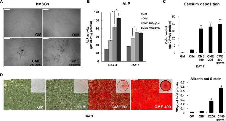
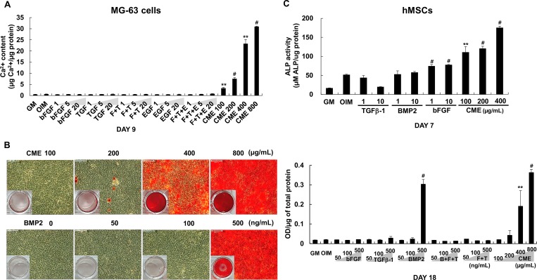
Human amniotic membrane extracts contain numerous growth factors and bioactive substances. However, osteogenic effects of amnion and chorion membrane extracts (AME and CME, respectively) on osteoblasts are unclear. In this study, we explored the ability of AME and CME to promote the osteogenic differentiation of osteoblast-like MG-63 cells. MG-63 cells were cultured in osteogenic induction medium (OIM) with or without exogenous AME and CME. CME enhanced the osteogenic differentiation of MG-63 cells compared with AME, as indicated by increased mineralization; alkaline phosphatase activity; and mRNA expression of osteogenic marker genes encoding integrin-binding sialoprotein (IBSP), RUNX2, OSTERIX, and osteocalcin (OCN). Interestingly, AME and CME contained different combinations of osteogenesis-related growth factors, including basic fibroblast growth factor (bFGF), transforming growth factor beta-1 (TGFβ-1), and epidermal growth factor (EGF), which differentially regulated the osteogenic differentiation of MG-63 cells. bFGF and TGFβ-1 present in CME positively regulated the osteogenic differentiation of MG-63 cells, whereas EGF present in AME negatively regulated the differentiation of MG-63 cells. Moreover, exogenous treatment of EGF antagonized CME-induced mineralization of extracellular matrix on MG-63 cells. We compared the osteogenic efficacy of CME with that of BMP2, bFGF, and TGFβ-1 alone or their combinations. We observed that CME greatly enhanced osteogenesis by providing a conductive environment for the differentiation of MG-63 cells. Together, our results indicated that human AME and CME exerted differential effects on osteogenesis because of the presence of different compositions of growth factors. In addition, our results highlighted a new possible strategy of using CME as a biocompatible therapeutic material for bone regeneration.
人羊膜提取物含有多种生长因子和生物活性物质。然而,羊膜和绒毛膜提取物(分别为AME和CME)对成骨细胞的成骨作用尚不清楚。在本研究中,我们探讨了AME和CME促进成骨样MG-63细胞成骨分化的能力。MG-63细胞在添加或不添加外源性AME和CME的成骨诱导培养基(OIM)中培养。与AME相比,CME增强了MG-63细胞的成骨分化,表现为矿化增加、碱性磷酸酶活性升高以及编码整合素结合唾液蛋白(IBSP)、RUNX2、osterix和骨钙素(OCN)的成骨标记基因的mRNA表达增加。有趣的是,AME和CME含有不同组合的成骨相关生长因子,包括碱性成纤维细胞生长因子(bFGF)、转化生长因子β-1(TGFβ-1)和表皮生长因子(EGF),它们对MG-63细胞的成骨分化有不同的调节作用。CME中存在的bFGF和TGFβ-1对MG-63细胞的成骨分化起正向调节作用,而AME中存在的EGF对MG-63细胞的分化起负向调节作用。此外,外源性EGF处理可拮抗CME诱导的MG-63细胞细胞外基质矿化。我们比较了CME与单独使用BMP2、bFGF和TGFβ-1或它们的组合的成骨效果。我们观察到CME通过为MG-63细胞的分化提供一个传导性环境,极大地增强了成骨作用。总之,我们的结果表明,由于生长因子组成不同,人AME和CME对成骨作用产生了不同的影响。此外,我们的结果突出了一种新的可能策略,即使用CME作为骨再生的生物相容性治疗材料。