Department of Gastroenterology and Hepatology, Nagasaki University Hospital, Nagasaki City, Nagasaki, Japan.
Department of Gastroenterology and Hepatology, Nagasaki University Graduate School of Biomedical Sciences, Nagasaki City, Nagasaki, Japan.
Med Sci Monit. 2017 Sep 21;23:4526-4532. doi: 10.12659/msm.903210.
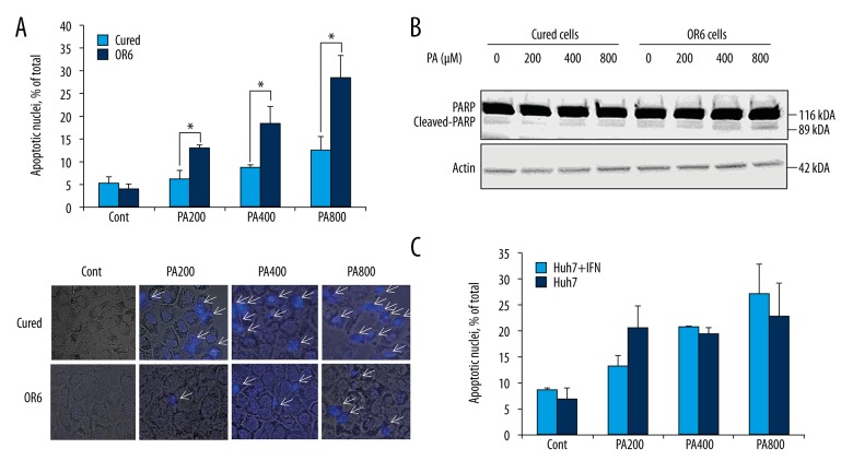
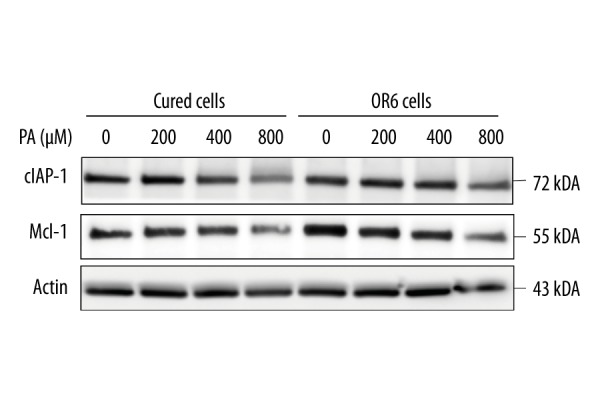
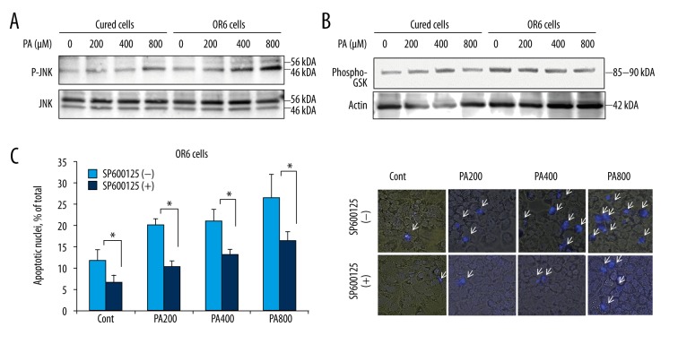
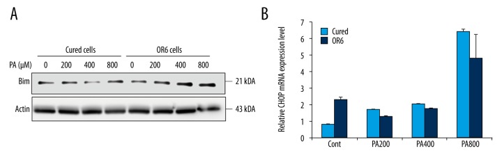
BACKGROUND Hepatitis C virus (HCV) infection and metabolic diseases including nonalcoholic steatohepatitis (NASH) exhibit a complex interplay. Although free fatty acid-mediated apoptosis is a prominent feature of NASH, the impact of HCV infection on hepatocyte lipotoxicity has remained largely unexplored. The study aimed at identifying whether infection by HCV affected the apoptotic pathway in hepatocytes during fatty acid assault. MATERIAL AND METHODS OR6 cells, which are derived from human hepatocellular carcinoma Huh-7 cells and harbor a full-length HCV RNA genome replication system, were treated with palmitate. Apoptosis was examined by 4',6-diamidino-2-phenylindole staining. Activation and expression of JNK, Bim, cIAP-1, and Mcl-1 were examined by immunoblotting. mRNA expression of CHOP, a major player in endoplasmic reticulum stress-mediated apoptosis, was assessed by real-time PCR. RESULTS Palmitate-induced hepatocyte apoptosis was significantly enhanced in OR6 cells compared to cured cells, in which the HCV genome had been eradicated by treatment with interferon-α. Although basal expression of CHOP mRNA was enhanced in OR6 cells compared to cured cells, it was similarly upregulated in both cell lines following palmitate treatment. Notably, palmitate-induced JNK phosphorylation was accentuated in OR6 cells compared to cured cells. Inhibition of JNK with SP600125 attenuated palmitate-induced apoptosis. Palmitate-mediated upregulation of BH3-only protein Bim, which acts downstream of JNK, was also enhanced in OR6 cells compared to cured cells. In contrast, Mcl-1 and cIAP-1 were equally reduced in OR6 cells and cured cells following palmitate treatment. CONCLUSIONS These findings suggest that during lipoapoptosis, HCV infection may enhance hepatocyte toxicity by increasing JNK phosphorylation.
丙型肝炎病毒(HCV)感染和非酒精性脂肪性肝炎(NASH)等代谢疾病之间存在复杂的相互作用。虽然游离脂肪酸介导的细胞凋亡是 NASH 的一个突出特征,但 HCV 感染对肝细胞脂毒性的影响在很大程度上仍未得到探索。本研究旨在确定 HCV 感染是否会影响脂肪酸攻击时肝细胞中的凋亡途径。
OR6 细胞源自人肝癌 Huh-7 细胞,携带全长 HCV RNA 基因组复制系统,用棕榈酸处理。通过 4',6-二脒基-2-苯基吲哚染色检查细胞凋亡。通过免疫印迹法检查 JNK、Bim、cIAP-1 和 Mcl-1 的激活和表达。通过实时 PCR 评估内质网应激介导的凋亡的主要参与者 CHOP 的 mRNA 表达。
与 cured 细胞相比,OR6 细胞中棕榈酸诱导的肝细胞凋亡明显增强,在 cured 细胞中,HCV 基因组已被干扰素-α治疗消除。与 cured 细胞相比,OR6 细胞中 CHOP mRNA 的基础表达增强,但在两种细胞系中,在用棕榈酸处理后,其表达均上调。值得注意的是,与 cured 细胞相比,OR6 细胞中棕榈酸诱导的 JNK 磷酸化增强。用 SP600125 抑制 JNK 可减弱棕榈酸诱导的细胞凋亡。OR6 细胞中 BH3 仅蛋白 Bim 的表达上调,这是 JNK 的下游,与 cured 细胞相比也增强,而 Mcl-1 和 cIAP-1 在 OR6 细胞和 cured 细胞中用棕榈酸处理后均等量减少。
这些发现表明,在脂肪细胞凋亡过程中,HCV 感染可能通过增加 JNK 磷酸化来增强肝细胞毒性。