MRC-Laboratory of Molecular Biology, Francis Crick Avenue, Cambridge CB2 2QH, UK.
MRC-Laboratory of Molecular Biology, Francis Crick Avenue, Cambridge CB2 2QH, UK.
Curr Biol. 2017 Oct 9;27(19):2951-2962.e5. doi: 10.1016/j.cub.2017.07.047. Epub 2017 Sep 21.
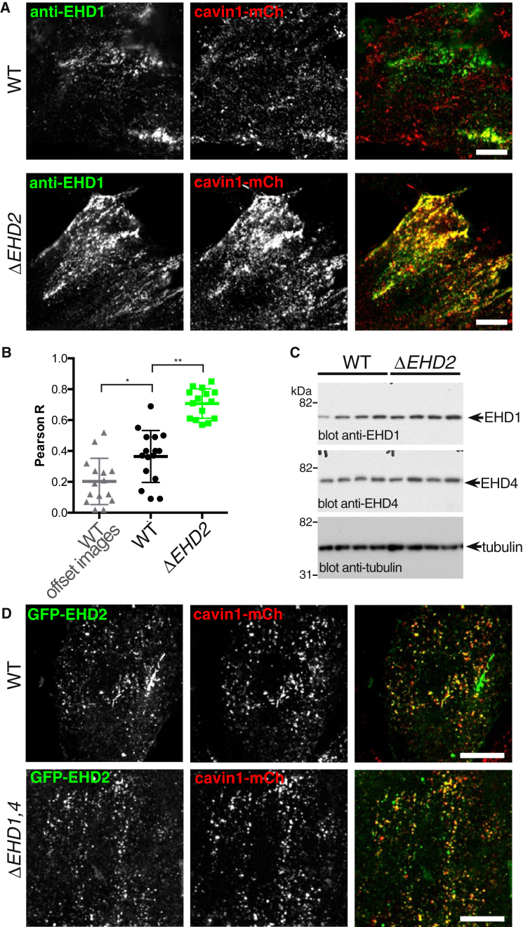
Caveolae introduce flask-shaped convolutions into the plasma membrane and help to protect the plasma membrane from damage under stretch forces. The protein components that form the bulb of caveolae are increasingly well characterized, but less is known about the contribution of proteins that localize to the constricted neck. Here we make extensive use of multiple CRISPR/Cas9-generated gene knockout and knockin cell lines to investigate the role of Eps15 Homology Domain (EHD) proteins at the neck of caveolae. We show that EHD1, EHD2, and EHD4 are recruited to caveolae. Recruitment of the other EHDs increases markedly when EHD2, which has been previously detected at caveolae, is absent. Construction of knockout cell lines lacking EHDs 1, 2, and 4 confirms this apparent functional redundancy. Two striking sets of phenotypes are observed in EHD1,2,4 knockout cells: (1) the characteristic clustering of caveolae into higher-order assemblies is absent; and (2) when the EHD1,2,4 knockout cells are subjected to prolonged cycles of stretch forces, caveolae are destabilized and the plasma membrane is prone to rupture. Our data identify the first molecular components that act to cluster caveolae into a membrane ultrastructure with the potential to extend stretch-buffering capacity and support a revised model for the function of EHDs at the caveolar neck.
陷窝小体在质膜上引入烧瓶状的卷曲,有助于保护质膜免受拉伸力的破坏。形成陷窝小体球部的蛋白成分越来越被充分描述,但对于定位于缩窄颈部的蛋白的贡献知之甚少。在这里,我们广泛使用多种 CRISPR/Cas9 生成的基因敲除和敲入细胞系来研究陷窝小体颈部 Eps15 同源结构域(EHD)蛋白的作用。我们表明,EHD1、EHD2 和 EHD4 被招募到陷窝小体。当以前在陷窝小体中检测到的 EHD2 不存在时,其他 EHD 的募集显著增加。敲除细胞系缺乏 EHDs 1、2 和 4 的构建证实了这种明显的功能冗余。在 EHD1、2、4 敲除细胞中观察到两组显著的表型:(1)陷窝小体特征性的聚集成更高阶的组装体不存在;(2)当 EHD1、2、4 敲除细胞经历长时间的拉伸力循环时,陷窝小体不稳定,质膜容易破裂。我们的数据确定了第一个分子成分,它们将陷窝小体聚集到具有潜在扩展拉伸缓冲能力的膜超结构中,并支持 EHD 在陷窝颈部功能的修正模型。