• 文献检索
  • 文档翻译
  • 深度研究
  • 学术资讯
  • Suppr Zotero 插件Zotero 插件
  • 邀请有礼
  • 套餐&价格
  • 历史记录
应用&插件
Suppr Zotero 插件Zotero 插件浏览器插件Mac 客户端Windows 客户端微信小程序
定价
高级版会员购买积分包购买API积分包
服务
文献检索文档翻译深度研究API 文档MCP 服务
关于我们
关于 Suppr公司介绍联系我们用户协议隐私条款
关注我们

Suppr 超能文献

核心技术专利:CN118964589B侵权必究
粤ICP备2023148730 号-1Suppr @ 2026

文献检索

告别复杂PubMed语法,用中文像聊天一样搜索,搜遍4000万医学文献。AI智能推荐,让科研检索更轻松。

立即免费搜索

文件翻译

保留排版,准确专业,支持PDF/Word/PPT等文件格式,支持 12+语言互译。

免费翻译文档

深度研究

AI帮你快速写综述,25分钟生成高质量综述,智能提取关键信息,辅助科研写作。

立即免费体验

采用独立成分分析对颈动脉狭窄患者进行数字减影血管造影的自动血流分析。

Automatic flow analysis of digital subtraction angiography using independent component analysis in patients with carotid stenosis.

作者信息

Lee Han-Jui, Hong Jia-Sheng, Lin Chung-Jung, Kao Yi-Hsuan, Chang Feng-Chi, Luo Chao-Bao, Chu Wei-Fa

机构信息

Department of Radiology, Taipei Veterans General Hospital, Taipei, Taiwan.

School of Medicine, National Yang-Ming University, Taipei, Taiwan.

出版信息

PLoS One. 2017 Sep 26;12(9):e0185330. doi: 10.1371/journal.pone.0185330. eCollection 2017.

DOI:10.1371/journal.pone.0185330
PMID:28949999
原文链接:https://pmc.ncbi.nlm.nih.gov/articles/PMC5614569/
Abstract

PURPOSE

Current time-density curve analysis of digital subtraction angiography (DSA) provides intravascular flow information but requires manual vasculature selection. We developed an angiographic marker that represents cerebral perfusion by using automatic independent component analysis.

MATERIALS AND METHODS

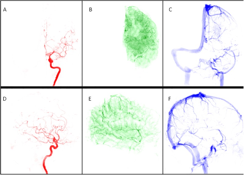
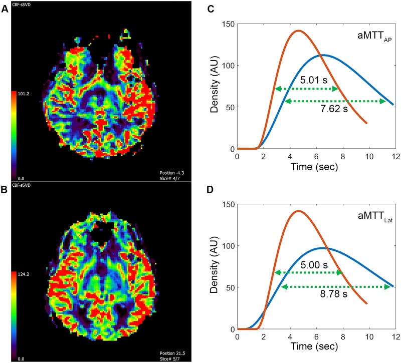
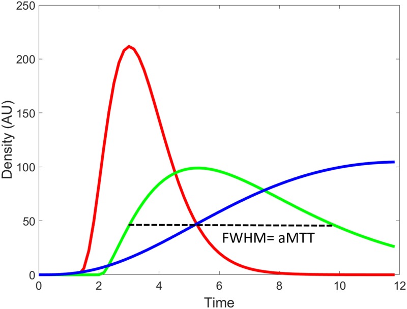
We retrospectively analyzed the data of 44 patients with unilateral carotid stenosis higher than 70% according to North American Symptomatic Carotid Endarterectomy Trial criteria. For all patients, magnetic resonance perfusion (MRP) was performed one day before DSA. Fixed contrast injection protocols and DSA acquisition parameters were used before stenting. The cerebral circulation time (CCT) was defined as the difference in the time to peak between the parietal vein and cavernous internal carotid artery in a lateral angiogram. Both anterior-posterior and lateral DSA views were processed using independent component analysis, and the capillary angiogram was extracted automatically. The full width at half maximum of the time-density curve in the capillary phase in the anterior-posterior and lateral DSA views was defined as the angiographic mean transient time (aMTT; i.e., aMTTAP and aMTTLat). The correlations between the degree of stenosis, CCT, aMTTAP and aMTTLat, and MRP parameters were evaluated.

RESULTS

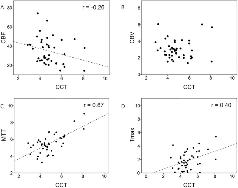
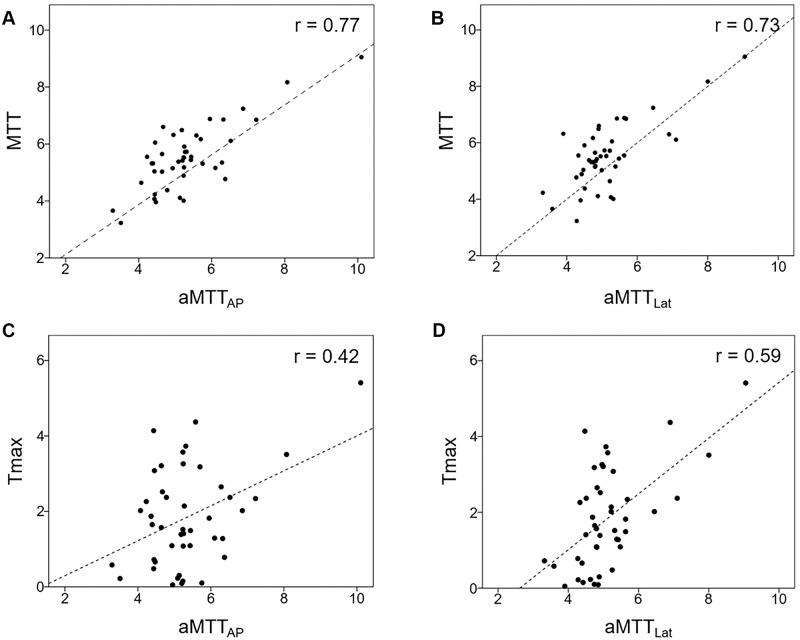
The degree of stenosis showed no correlation with CCT, aMTTAP, aMTTLat, or any MRP parameter. CCT showed a strong correlation with aMTTAP (r = 0.67) and aMTTLat (r = 0.72). Among the MRP parameters, CCT showed only a moderate correlation with MTT (r = 0.67) and Tmax (r = 0.40). aMTTAP showed a moderate correlation with Tmax (r = 0.42) and a strong correlation with MTT (r = 0.77). aMTTLat also showed similar correlations with Tmax (r = 0.59) and MTT (r = 0.73).

CONCLUSION

Apart from vascular anatomy, aMTT estimates brain parenchyma hemodynamics from DSA and is concordant with MRP. This process is completely automatic and provides immediate measurement of quantitative peritherapeutic brain parenchyma changes during stenting.

摘要

目的

目前数字减影血管造影(DSA)的时间密度曲线分析可提供血管内血流信息,但需要手动选择血管。我们通过使用自动独立成分分析开发了一种代表脑灌注的血管造影标记物。

材料与方法

我们根据北美症状性颈动脉内膜切除术试验标准,回顾性分析了44例单侧颈动脉狭窄高于70%患者的数据。对所有患者在DSA前一天进行磁共振灌注(MRP)检查。在支架置入前使用固定的造影剂注射方案和DSA采集参数。脑循环时间(CCT)定义为侧位血管造影中顶叶静脉与海绵窦段颈内动脉之间的峰值时间差。前后位和侧位DSA图像均使用独立成分分析进行处理,并自动提取毛细血管造影图像。前后位和侧位DSA图像中毛细血管期时间密度曲线的半高全宽定义为血管造影平均通过时间(aMTT;即aMTTAP和aMTTLat)。评估狭窄程度、CCT、aMTTAP和aMTTLat与MRP参数之间的相关性。

结果

狭窄程度与CCT、aMTTAP、aMTTLat或任何MRP参数均无相关性。CCT与aMTTAP(r = 0.67)和aMTTLat(r = 0.72)呈强相关性。在MRP参数中,CCT仅与MTT(r = 0.67)和Tmax(r = 0.40)呈中度相关性。aMTTAP与Tmax(r = 0.42)呈中度相关性,与MTT(r = 0.77)呈强相关性。aMTTLat与Tmax(r = 0.59)和MTT(r = 0.73)也呈相似的相关性。

结论

除血管解剖结构外,aMTT可从DSA估计脑实质血流动力学,且与MRP结果一致。该过程完全自动化,并可在支架置入期间即时测量治疗期间脑实质的定量变化。

https://cdn.ncbi.nlm.nih.gov/pmc/blobs/b4f0/5614569/5e42096c5083/pone.0185330.g005.jpg
https://cdn.ncbi.nlm.nih.gov/pmc/blobs/b4f0/5614569/bf557dffa5c3/pone.0185330.g001.jpg
https://cdn.ncbi.nlm.nih.gov/pmc/blobs/b4f0/5614569/bbfdc66667fc/pone.0185330.g002.jpg
https://cdn.ncbi.nlm.nih.gov/pmc/blobs/b4f0/5614569/a13ca15d4629/pone.0185330.g003.jpg
https://cdn.ncbi.nlm.nih.gov/pmc/blobs/b4f0/5614569/0219fe2ce959/pone.0185330.g004.jpg
https://cdn.ncbi.nlm.nih.gov/pmc/blobs/b4f0/5614569/5e42096c5083/pone.0185330.g005.jpg

相似文献

1
Automatic flow analysis of digital subtraction angiography using independent component analysis in patients with carotid stenosis.采用独立成分分析对颈动脉狭窄患者进行数字减影血管造影的自动血流分析。
PLoS One. 2017 Sep 26;12(9):e0185330. doi: 10.1371/journal.pone.0185330. eCollection 2017.
2
Evaluating cerebral hemodynamics using quantitative digital subtraction angiography and flat-detector computed tomography perfusion imaging: A comparative study in patients with carotid stenosis.采用定量数字减影血管造影和平板探测器 CT 灌注成像评估脑血流动力学:颈动脉狭窄患者的对比研究。
J Chin Med Assoc. 2018 Nov;81(11):977-984. doi: 10.1016/j.jcma.2018.01.016. Epub 2018 Aug 23.
3
Finding the optimal deconvolution algorithm for MR perfusion in carotid stenosis: Correlations with angiographic cerebral circulation time.寻找颈动脉狭窄磁共振灌注的最佳去卷积算法:与血管造影脑循环时间的相关性
J Neuroradiol. 2016 Jul;43(4):290-6. doi: 10.1016/j.neurad.2016.02.002. Epub 2016 Mar 30.
4
Fluoroscopic angiography quantifies delay in cerebral circulation time and requires less radiation in carotid stenosis patients: A pilot study.荧光透视血管造影定量评估了脑循环时间的延迟,并且在颈动脉狭窄患者中需要的辐射更少:一项初步研究。
J Chin Med Assoc. 2019 May;82(5):396-400. doi: 10.1097/JCMA.0000000000000046.
5
Prolonged cerebral circulation time is more associated with symptomatic carotid stenosis than stenosis degree or collateral circulation.大脑循环时间延长与症状性颈动脉狭窄的相关性高于狭窄程度或侧支循环。
J Neurointerv Surg. 2018 May;10(5):476-480. doi: 10.1136/neurintsurg-2017-013293. Epub 2017 Aug 30.
6
Validating the Automatic Independent Component Analysis of DSA.验证 DSA 的自动独立成分分析。
AJNR Am J Neuroradiol. 2019 Mar;40(3):540-542. doi: 10.3174/ajnr.A5963. Epub 2019 Jan 24.
7
Changes of time-attenuation curve blood flow parameters in patients with and without carotid stenosis.有和没有颈动脉狭窄的患者时间-衰减曲线血流参数的变化。
AJNR Am J Neuroradiol. 2015 Jun;36(6):1176-81. doi: 10.3174/ajnr.A4239. Epub 2015 Feb 26.
8
Correlation of Computed Tomography Angiography to Digital Subtraction Angiography in Carotid Stenosis with Real-World Assessment of Overestimation of Carotid Stenosis on Computed Tomography Angiography.基于真实世界评估的 CT 血管造影对颈动脉狭窄高估的相关性研究与数字减影血管造影术在颈动脉狭窄中的应用。
World Neurosurg. 2024 Sep;189:e452-e458. doi: 10.1016/j.wneu.2024.06.084. Epub 2024 Jun 19.
9
Internal carotid artery stenosis measurement: comparison of 3D computed rotational angiography and conventional digital subtraction angiography.颈内动脉狭窄测量:三维计算机旋转血管造影与传统数字减影血管造影的比较
Stroke. 2004 Dec;35(12):2776-81. doi: 10.1161/01.STR.0000147037.12223.d5. Epub 2004 Oct 28.
10
Measurement of carotid stenosis using duplex ultrasonography with a microconvex array transducer: a validation with cerebral angiography.应用微凸阵探头的双功能超声检测颈动脉狭窄:与脑血管造影术的对照验证。
J Stroke Cerebrovasc Dis. 2013 Nov;22(8):e360-5. doi: 10.1016/j.jstrokecerebrovasdis.2013.03.028. Epub 2013 Apr 20.

引用本文的文献

1
perfDSA: Automatic Perfusion Imaging in Cerebral Digital Subtraction Angiography.灌注数字减影血管造影:脑数字减影血管造影中的自动灌注成像
Int J Comput Assist Radiol Surg. 2025 Apr 24. doi: 10.1007/s11548-025-03359-4.
2
Spatiotemporal Disentanglement of Arteriovenous Malformations in Digital Subtraction Angiography.数字减影血管造影中动静脉畸形的时空解缠
Proc SPIE Int Soc Opt Eng. 2024 Feb;12926. doi: 10.1117/12.3006740. Epub 2024 Apr 2.
3
CFD Study of the Effect of the Angle Pattern on Iliac Vein Compression Syndrome.髂静脉压迫综合征中角度模式影响的计算流体动力学研究

本文引用的文献

1
Quantifying the Cerebral Hemodynamics of Dural Arteriovenous Fistula in Transverse Sigmoid Sinus Complicated by Sinus Stenosis: A Retrospective Cohort Study.量化横窦乙状窦窦狭窄合并硬脑膜动静脉瘘的脑血流动力学:一项回顾性队列研究。
AJNR Am J Neuroradiol. 2017 Jan;38(1):132-138. doi: 10.3174/ajnr.A4960. Epub 2016 Oct 20.
2
Perfusion Angiography in Acute Ischemic Stroke.急性缺血性卒中的灌注血管造影
Comput Math Methods Med. 2016;2016:2478324. doi: 10.1155/2016/2478324. Epub 2016 Jul 3.
3
Using Flat-Panel Perfusion Imaging to Measure Cerebral Hemodynamics: A Pilot Feasibility Study in Patients With Carotid Stenosis.
Bioengineering (Basel). 2023 Jun 5;10(6):688. doi: 10.3390/bioengineering10060688.
4
Peri-therapeutic multi-modal hemodynamic assessment and detection of predictors for symptomatic in-stent restenosis after percutaneous transluminal angioplasty and stenting.经皮腔内血管成形术和支架置入术后治疗期间的多模态血流动力学评估及有症状支架内再狭窄预测因素的检测
Front Neurol. 2023 Apr 18;14:1136847. doi: 10.3389/fneur.2023.1136847. eCollection 2023.
5
Feasibility of a silicone vascular phantom replicating real arterial contrast filling dynamics on cerebral angiography: An in-vitro pilot study.硅酮血管模型在脑血管造影中模拟真实动脉对比剂充盈动力学的可行性:一项体外初步研究。
PLoS One. 2023 Jan 17;18(1):e0280395. doi: 10.1371/journal.pone.0280395. eCollection 2023.
6
Carotid webs produce greater hemodynamic disturbances than atherosclerotic disease: a DSA time-density curve study.颈动脉梳状结构比动脉粥样硬化性疾病产生更大的血液动力学紊乱:一项 DSA 时间密度曲线研究。
J Neurointerv Surg. 2022 Jul;14(7):729-733. doi: 10.1136/neurintsurg-2021-017588. Epub 2021 Jul 27.
7
Quantitative Evaluation of Peripheral Arterial Blood Flow Using Peri-Interventional Fluoroscopic Parameters: An In Vivo Study Evaluating Feasibility and Clinical Utility.应用介入术中透视参数对周围动脉血流进行定量评估:一项评估可行性和临床应用价值的体内研究。
Biomed Res Int. 2020 Jan 16;2020:9526790. doi: 10.1155/2020/9526790. eCollection 2020.
使用平板灌注成像测量脑血流动力学:一项针对颈动脉狭窄患者的初步可行性研究。
Medicine (Baltimore). 2016 May;95(20):e3529. doi: 10.1097/MD.0000000000003529.
4
Peritherapeutic Hemodynamic Changes of Carotid Stenting Evaluated with Quantitative DSA in Patients with Carotid Stenosis.应用定量数字减影血管造影术评估颈动脉狭窄患者颈动脉支架置入术围治疗期血流动力学变化
AJNR Am J Neuroradiol. 2016 Oct;37(10):1883-1888. doi: 10.3174/ajnr.A4813. Epub 2016 May 12.
5
Finding the optimal deconvolution algorithm for MR perfusion in carotid stenosis: Correlations with angiographic cerebral circulation time.寻找颈动脉狭窄磁共振灌注的最佳去卷积算法:与血管造影脑循环时间的相关性
J Neuroradiol. 2016 Jul;43(4):290-6. doi: 10.1016/j.neurad.2016.02.002. Epub 2016 Mar 30.
6
Compensatory patterns of collateral flow in stroke patients with unilateral and bilateral carotid stenosis.单侧和双侧颈动脉狭窄的中风患者侧支血流的代偿模式。
BMC Neurol. 2016 Mar 18;16:39. doi: 10.1186/s12883-016-0560-0.
7
Dynamic Angiography and Perfusion Imaging Using Flat Detector CT in the Angiography Suite: A Pilot Study in Patients with Acute Middle Cerebral Artery Occlusions.在血管造影套件中使用平板探测器CT进行动态血管造影和灌注成像:急性大脑中动脉闭塞患者的初步研究。
AJNR Am J Neuroradiol. 2015 Oct;36(10):1964-70. doi: 10.3174/ajnr.A4383. Epub 2015 Jun 11.
8
Effective dose to patient measurements in flat-detector and multislice computed tomography: a comparison of applications in neuroradiology.在平板探测器和多层 CT 中对患者进行有效剂量测量:在神经放射学中的应用比较。
Eur Radiol. 2014 Jun;24(6):1257-65. doi: 10.1007/s00330-014-3136-7. Epub 2014 Apr 2.
9
Stenotic transverse sinus predisposes to poststenting hyperperfusion syndrome as evidenced by quantitative analysis of peritherapeutic cerebral circulation time.狭窄的横窦易引发支架置入术后高灌注综合征,治疗期间脑循环时间的定量分析证实了这一点。
AJNR Am J Neuroradiol. 2014 Jun;35(6):1132-6. doi: 10.3174/ajnr.A3838. Epub 2014 Jan 16.
10
Arterial input function in perfusion MRI: a comprehensive review.灌注 MRI 的动脉输入函数:全面综述。
Prog Nucl Magn Reson Spectrosc. 2013 Oct;74:1-32. doi: 10.1016/j.pnmrs.2013.04.002. Epub 2013 May 11.