• 文献检索
  • 文档翻译
  • 深度研究
  • 学术资讯
  • Suppr Zotero 插件Zotero 插件
  • 邀请有礼
  • 套餐&价格
  • 历史记录
应用&插件
Suppr Zotero 插件Zotero 插件浏览器插件Mac 客户端Windows 客户端微信小程序
定价
高级版会员购买积分包购买API积分包
服务
文献检索文档翻译深度研究API 文档MCP 服务
关于我们
关于 Suppr公司介绍联系我们用户协议隐私条款
关注我们

Suppr 超能文献

核心技术专利:CN118964589B侵权必究
粤ICP备2023148730 号-1Suppr @ 2026

文献检索

告别复杂PubMed语法,用中文像聊天一样搜索,搜遍4000万医学文献。AI智能推荐,让科研检索更轻松。

立即免费搜索

文件翻译

保留排版,准确专业,支持PDF/Word/PPT等文件格式,支持 12+语言互译。

免费翻译文档

深度研究

AI帮你快速写综述,25分钟生成高质量综述,智能提取关键信息,辅助科研写作。

立即免费体验

冠状动脉内成像在急性冠状动脉综合征中的作用:聚焦光学相干断层扫描

The role of intracoronary imaging in acute coronary syndromes: OCT in focus.

作者信息

Samir Ahmad, ElGuindy Ahmed

机构信息

Aswan Heart Centre, Aswan, Egypt.

Faculty of Medicine, Cairo University, Egypt.

出版信息

Glob Cardiol Sci Pract. 2016 Dec 30;2016(4):e201636. doi: 10.21542/gcsp.2016.36.

DOI:10.21542/gcsp.2016.36
PMID:28979905
原文链接:https://pmc.ncbi.nlm.nih.gov/articles/PMC5624192/
Abstract

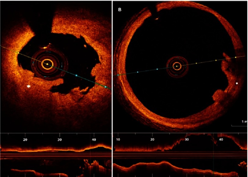
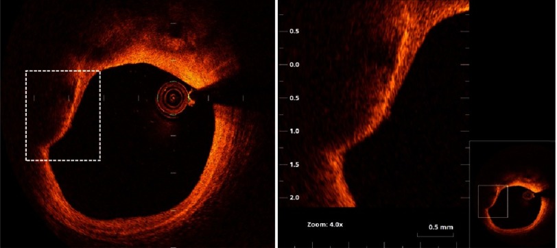
Optical coherence tomography (OCT) has emerged as a powerful intravascular imaging modality in recent years. The introduction of frequency-domain OCT has simplified the procedure and enabled its safe utilisation in different clinical settings including acute coronary syndromes, where it can determine the mechanism of plaque disruption, thrombus burden, and guide percutaneous coronary intervention. In patients presenting with stent failure (stent thrombosis and instent restenosis), OCT can also be very useful in determining the underlying mechanism and guiding therapy thereafter. This article aims to review the role of OCT in acute coronary syndromes as well as its potential clinical applications.

摘要

近年来,光学相干断层扫描(OCT)已成为一种强大的血管内成像方式。频域OCT的引入简化了操作流程,并使其能够安全应用于包括急性冠状动脉综合征在内的不同临床场景,在这些场景中,它可以确定斑块破裂的机制、血栓负荷,并指导经皮冠状动脉介入治疗。对于出现支架故障(支架血栓形成和支架内再狭窄)的患者,OCT在确定潜在机制并指导后续治疗方面也非常有用。本文旨在综述OCT在急性冠状动脉综合征中的作用及其潜在的临床应用。

https://cdn.ncbi.nlm.nih.gov/pmc/blobs/1d98/5624192/7ea9d58cf109/gcsp-2016-4-e201636-g012.jpg
https://cdn.ncbi.nlm.nih.gov/pmc/blobs/1d98/5624192/a90c29d41838/gcsp-2016-4-e201636-g001.jpg
https://cdn.ncbi.nlm.nih.gov/pmc/blobs/1d98/5624192/994fef7efaf4/gcsp-2016-4-e201636-g002.jpg
https://cdn.ncbi.nlm.nih.gov/pmc/blobs/1d98/5624192/31a0ee769316/gcsp-2016-4-e201636-g003.jpg
https://cdn.ncbi.nlm.nih.gov/pmc/blobs/1d98/5624192/527271d68447/gcsp-2016-4-e201636-g004.jpg
https://cdn.ncbi.nlm.nih.gov/pmc/blobs/1d98/5624192/33068d08dd69/gcsp-2016-4-e201636-g005.jpg
https://cdn.ncbi.nlm.nih.gov/pmc/blobs/1d98/5624192/966b185a2481/gcsp-2016-4-e201636-g006.jpg
https://cdn.ncbi.nlm.nih.gov/pmc/blobs/1d98/5624192/12aac9a8f720/gcsp-2016-4-e201636-g007.jpg
https://cdn.ncbi.nlm.nih.gov/pmc/blobs/1d98/5624192/528cc1ded040/gcsp-2016-4-e201636-g008.jpg
https://cdn.ncbi.nlm.nih.gov/pmc/blobs/1d98/5624192/4989f6bc8ff5/gcsp-2016-4-e201636-g009.jpg
https://cdn.ncbi.nlm.nih.gov/pmc/blobs/1d98/5624192/d6f9fef42fbc/gcsp-2016-4-e201636-g010.jpg
https://cdn.ncbi.nlm.nih.gov/pmc/blobs/1d98/5624192/8d8224d9e60d/gcsp-2016-4-e201636-g011.jpg
https://cdn.ncbi.nlm.nih.gov/pmc/blobs/1d98/5624192/7ea9d58cf109/gcsp-2016-4-e201636-g012.jpg

相似文献

1
The role of intracoronary imaging in acute coronary syndromes: OCT in focus.冠状动脉内成像在急性冠状动脉综合征中的作用:聚焦光学相干断层扫描
Glob Cardiol Sci Pract. 2016 Dec 30;2016(4):e201636. doi: 10.21542/gcsp.2016.36.
2
The diagnostic value of intracoronary optical coherence tomography.冠状动脉内光学相干断层扫描的诊断价值
Herz. 2011 Aug;36(5):417-29. doi: 10.1007/s00059-011-3487-7.
3
The Role of Intracoronary Plaque Imaging with Intravascular Ultrasound, Optical Coherence Tomography, and Near-Infrared Spectroscopy in Patients with Coronary Artery Disease.冠状动脉内斑块成像(血管内超声、光学相干断层扫描和近红外光谱)在冠状动脉疾病患者中的作用。
Curr Atheroscler Rep. 2016 Sep;18(9):57. doi: 10.1007/s11883-016-0607-0.
4
Optical coherence tomography: from research to clinical application.光学相干断层扫描:从研究到临床应用。
Minerva Med. 2012 Dec;103(6):441-64.
5
ST-segment elevation myocardial infarction with plaque erosion, to stent or not to stent: utility of intracoronary optical coherence tomography (OCT) imaging-a case report.伴有斑块侵蚀的ST段抬高型心肌梗死,是否植入支架:冠状动脉光学相干断层扫描(OCT)成像的效用——病例报告
Eur Heart J Case Rep. 2022 Feb 15;6(2):ytac078. doi: 10.1093/ehjcr/ytac078. eCollection 2022 Feb.
6
Optical Coherence Tomography Guidance In The Management Of Acute Coronary Syndrome Based On Plaque Morphology.光学相干断层扫描指导基于斑块形态的急性冠状动脉综合征管理。
J Ayub Med Coll Abbottabad. 2021 Jan-Mar;33(1):26-29.
7
Clinical Characterization of Coronary Atherosclerosis With Dual-Modality OCT and Near-Infrared Autofluorescence Imaging.冠状动脉粥样硬化的双模态光学相干断层扫描和近红外自发荧光成像的临床特征
JACC Cardiovasc Imaging. 2016 Nov;9(11):1304-1314. doi: 10.1016/j.jcmg.2015.11.020. Epub 2016 Mar 9.
8
Optical Coherence Tomography in Intracoronary Diagnostics.
S D Med. 2020 May;73(5):202-207.
9
Combined use of optical coherence tomography and intravascular ultrasound imaging in patients undergoing coronary interventions for stent thrombosis.光学相干断层扫描和血管内超声成像在冠状动脉介入治疗支架血栓形成患者中的联合应用。
Heart. 2012 Aug;98(16):1213-20. doi: 10.1136/heartjnl-2012-302183.
10
Feasibility and repeatability of optical coherence tomography measurements of pre-stent thrombus burden in patients with STEMI treated with primary PCI.光学相干断层扫描测量 STEMI 患者直接 PCI 治疗后支架内血栓负荷的可行性和可重复性。
Eur Heart J Cardiovasc Imaging. 2015 Jan;16(1):96-107. doi: 10.1093/ehjci/jeu175. Epub 2014 Sep 19.

引用本文的文献

1
Optical Coherence Tomography in Myocardial Infarction Management: Enhancing Precision in Percutaneous Coronary Intervention.光学相干断层扫描在心肌梗死管理中的应用:提高经皮冠状动脉介入治疗的精准度
J Clin Med. 2024 Sep 28;13(19):5791. doi: 10.3390/jcm13195791.
2
What eyes do not see the heart does not grieve over? The role of intracoronary imaging in acute myocardial infarction: a case report.眼不见心不烦?冠状动脉内成像在急性心肌梗死中的作用:一例报告
Eur Heart J Case Rep. 2023 Sep 8;7(9):ytad444. doi: 10.1093/ehjcr/ytad444. eCollection 2023 Sep.
3
Culprit Lesion Morphology on Optical Coherence Tomography in ST-elevation Myocardial Infarction vs Non ST-elevation Myocardial Infarction - A Systematic Review of 7526 Patients.

本文引用的文献

1
Optical Coherence Tomography to Optimize Results of Percutaneous Coronary Intervention in Patients with Non-ST-Elevation Acute Coronary Syndrome: Results of the Multicenter, Randomized DOCTORS Study (Does Optical Coherence Tomography Optimize Results of Stenting).光学相干断层成像术优化非 ST 段抬高型急性冠状动脉综合征患者经皮冠状动脉介入治疗的结果:多中心、随机 DOCTORS 研究的结果(光学相干断层成像术是否优化支架置入术的结果)。
Circulation. 2016 Sep 27;134(13):906-17. doi: 10.1161/CIRCULATIONAHA.116.024393. Epub 2016 Aug 29.
2
Coronary stent thrombosis: what have we learned?冠状动脉支架血栓形成:我们学到了什么?
J Thorac Dis. 2016 Jul;8(7):1398-405. doi: 10.21037/jtd.2016.05.69.
3
ST段抬高型心肌梗死与非ST段抬高型心肌梗死光学相干断层扫描的罪犯病变形态学——对7526例患者的系统评价
J Saudi Heart Assoc. 2023 Mar 9;35(1):40-49. doi: 10.37616/2212-5043.1329. eCollection 2023.
High-sensitivity intravascular photoacoustic imaging of lipid-laden plaque with a collinear catheter design.
采用共线导管设计对富含脂质斑块进行高灵敏度血管内光声成像。
Sci Rep. 2016 Apr 28;6:25236. doi: 10.1038/srep25236.
4
Mechanisms of stent thrombosis analysed by optical coherence tomography: insights from the national PESTO French registry.光学相干断层成像术分析支架内血栓形成的机制:来自法国全国 PESTO 登记研究的见解。
Eur Heart J. 2016 Apr 14;37(15):1208-16. doi: 10.1093/eurheartj/ehv711. Epub 2016 Jan 12.
5
Stent thrombosis and restenosis: what have we learned and where are we going? The Andreas Grüntzig Lecture ESC 2014.支架内血栓形成与再狭窄:我们学到了什么,又将何去何从?2014年欧洲心脏病学会安德烈亚斯·格鲁恩齐格讲座
Eur Heart J. 2015 Dec 14;36(47):3320-31. doi: 10.1093/eurheartj/ehv511. Epub 2015 Sep 28.
6
Optical coherence tomography imaging during percutaneous coronary intervention impacts physician decision-making: ILUMIEN I study.经皮冠状动脉介入治疗期间的光学相干断层扫描成像影响医生决策:ILUMIEN I研究。
Eur Heart J. 2015 Dec 14;36(47):3346-55. doi: 10.1093/eurheartj/ehv367. Epub 2015 Aug 4.
7
Suboptimal stent deployment is associated with subacute stent thrombosis: optical coherence tomography insights from a multicenter matched study. From the CLI Foundation investigators: the CLI-THRO study.支架植入不理想与亚急性支架血栓形成相关:一项多中心配对研究的光学相干断层扫描见解。CLI基金会研究人员:CLI-THRO研究。
Am Heart J. 2015 Feb;169(2):249-56. doi: 10.1016/j.ahj.2014.11.012. Epub 2014 Nov 20.
8
2014 ESC/EACTS Guidelines on myocardial revascularization: The Task Force on Myocardial Revascularization of the European Society of Cardiology (ESC) and the European Association for Cardio-Thoracic Surgery (EACTS)Developed with the special contribution of the European Association of Percutaneous Cardiovascular Interventions (EAPCI).2014年欧洲心脏病学会/欧洲心胸外科学会心肌血运重建指南:欧洲心脏病学会(ESC)和欧洲心胸外科学会(EACTS)心肌血运重建特别工作组编写,欧洲经皮心血管介入协会(EAPCI)提供特别贡献。
Eur Heart J. 2014 Oct 1;35(37):2541-619. doi: 10.1093/eurheartj/ehu278. Epub 2014 Aug 29.
9
We should use the OCT-based clinical term "acute coronary syndrome with intact fibrous cap (ACS-IFC)" rather than the pathology term "plaque erosion".
J Am Coll Cardiol. 2014 Jun 24;63(24):2745. doi: 10.1016/j.jacc.2013.09.087. Epub 2014 Apr 23.
10
Intravascular photoacoustic imaging: a new tool for vulnerable plaque identification.血管内光声成像:一种识别易损斑块的新工具。
Ultrasound Med Biol. 2014 Jun;40(6):1037-48. doi: 10.1016/j.ultrasmedbio.2014.01.008. Epub 2014 Mar 14.